

420
M. V. L. BENNETT
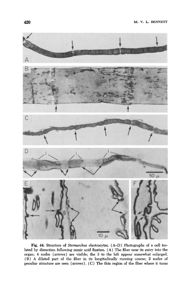
Fig. 44. Structure of Stemarchus electrocytes. (A-D) Photographs of a cell isolated by dissection following osmic acid fixation. ( A ) The fiber near its entry into the
organ; 4 nodes (arrows) are visible; the 3 to the left appear somewhat enlarged.
( B ) A dilated part of the fiber in its longitudinally running course; 2 nodes of
peculiar structure are seen (arrows). ( C ) The thin region of the fiber where it turns
M. V. L. BENNETT
Fig. 44. Structure of Stemarchus electrocytes. (A-D) Photographs of a cell isolated by dissection following osmic acid fixation. ( A ) The fiber near its entry into the
organ; 4 nodes (arrows) are visible; the 3 to the left appear somewhat enlarged.
( B ) A dilated part of the fiber in its longitudinally running course; 2 nodes of
peculiar structure are seen (arrows). ( C ) The thin region of the fiber where it turns