

10. ELECTRIC ORGANS
375
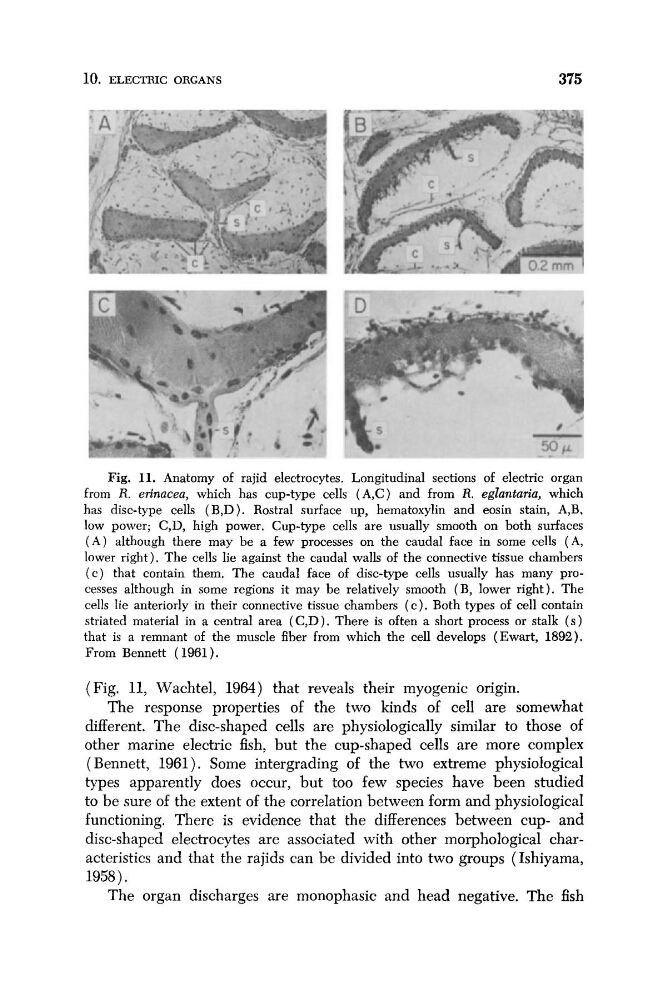
Fig. 11. Anatomy of rajid electrocytes. Longitudinal sections of electric organ
from R. erinacea, which has cup-type cells (A,C) and from R. eglanturia, which
has disc-type cells (B,D). Rostra1 surface up, hematoxylin and eosin stain, A,B,
low power; C,D, high power. Cup-type cells are usually smooth on both surfaces
( A ) although there may be a few processes on the caudal face in some cells (A,
lower right). The cells lie against the caudal walls of the connective tissue chambers
( c ) that contain them. The caudal face of disc-type cells usually has many processes although in some regions it may be relatively smooth (B, lower right). The
cells lie anteriorly in their connective tissue chambers ( c ) . Both types of cell contain
striated material in a central area (C,D). There is often a short process or stalk ( s )
that is a remnant of the muscle fiber from which the cell develops (Ewart, 1892).
From Bennett ( 1961).
(Fig. 11, Wachtel, 1964) that reveals their myogenic origin.
The response properties of the two kinds of cell are somewhat
different. The disc-shaped cells are physiologically similar to those of
other marine electric fish, but the cup-shaped cells are more complex
(Bennett, 1961). Some intergrading of the two extreme physiological
types apparently does occur, but too few species have been studied
to be sure of the extent of the correlation between form and physiological
functioning. There is evidence that the differences between cup- and
disc-shaped electrocytes are associated with other morphological characteristics and that the rajids can be divided into two groups (Ishiyama,
1958).
The organ discharges are monophasic and head negative. The fish
375
Fig. 11. Anatomy of rajid electrocytes. Longitudinal sections of electric organ
from R. erinacea, which has cup-type cells (A,C) and from R. eglanturia, which
has disc-type cells (B,D). Rostra1 surface up, hematoxylin and eosin stain, A,B,
low power; C,D, high power. Cup-type cells are usually smooth on both surfaces
( A ) although there may be a few processes on the caudal face in some cells (A,
lower right). The cells lie against the caudal walls of the connective tissue chambers
( c ) that contain them. The caudal face of disc-type cells usually has many processes although in some regions it may be relatively smooth (B, lower right). The
cells lie anteriorly in their connective tissue chambers ( c ) . Both types of cell contain
striated material in a central area (C,D). There is often a short process or stalk ( s )
that is a remnant of the muscle fiber from which the cell develops (Ewart, 1892).
From Bennett ( 1961).
(Fig. 11, Wachtel, 1964) that reveals their myogenic origin.
The response properties of the two kinds of cell are somewhat
different. The disc-shaped cells are physiologically similar to those of
other marine electric fish, but the cup-shaped cells are more complex
(Bennett, 1961). Some intergrading of the two extreme physiological
types apparently does occur, but too few species have been studied
to be sure of the extent of the correlation between form and physiological
functioning. There is evidence that the differences between cup- and
disc-shaped electrocytes are associated with other morphological characteristics and that the rajids can be divided into two groups (Ishiyama,
1958).
The organ discharges are monophasic and head negative. The fish