

366
M. V. L. BENNE'JT
_F
-A
C
D
F
H
D
B
-A E
H
G
-' +I;
I msec
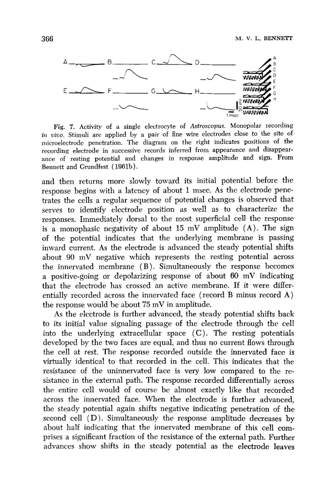
Fig. 7. Activity of a single electrocyte of Astroscopus. Monopolar recording
ill zjizjo. Stimuli are applied by a pair of fine wire electrodes close to the site of
microelectrode penetration. The diagram on the right indicates positions of the
recording electrode in successive records inferred from appearance and disappearance of resting potential and changes in response amplitude and sign. From
Bennett and Grundfest ( 1961b).
and then returns more slowly toward its initial potential before the
response begins with a latency of about 1 msec. As the electrode penetrates the cells a regular sequence of potential changes is observed that
serves to identify electrode position as well as to characterize the
responses. Immediately dorsal to the most superficial cell the response
is a monophasic negativity of about 15 mV amplitude ( A ) . The sign
of the potential indicates that the underlying membrane is passing
inward current. As the electrode is advanced the steady potential shifts
about 90 mV negative which represents the resting potential across
the innervated membrane ( B ) . Simultaneously the response becomes
a positive-going or depolarizing response of about 60 mV indicating
that the electrode has crossed an active membrane. If it were differentially recorded across the innervated face (record B minus record A )
the response would be about 75 mV in amplitude.
As the electrode is further advanced, the steady potential shifts back
to its initial value signaling passage of the electrode through the cell
into the underlying extracellular space ( C ) . The resting potentials
developed by the two faces are equal, and thus no current flows through
the cell at rest. The response recorded outside the innervated face is
virtually identical to that recorded in the cell. This indicates that the
resistance of the uninnervated face is very low compared to the resistance in the external path. The response recorded differentially across
the entire cell would of course be almost exactly like that recorded
across the innervated face. When the electrode is further advanced,
the steady potential again shifts negative indicating penetration of the
second cell ( D ) . Simultaneously the response amplitude decreases by
about half indicating that the innervated membrane of this cell cornprises a significant fraction of the resistance of the external path. Further
advances show shifts in the steady potential as the electrode leaves
M. V. L. BENNE'JT
_F
-A
C
D
F
H
D
B
-A E
H
G
-' +I;
I msec
Fig. 7. Activity of a single electrocyte of Astroscopus. Monopolar recording
ill zjizjo. Stimuli are applied by a pair of fine wire electrodes close to the site of
microelectrode penetration. The diagram on the right indicates positions of the
recording electrode in successive records inferred from appearance and disappearance of resting potential and changes in response amplitude and sign. From
Bennett and Grundfest ( 1961b).
and then returns more slowly toward its initial potential before the
response begins with a latency of about 1 msec. As the electrode penetrates the cells a regular sequence of potential changes is observed that
serves to identify electrode position as well as to characterize the
responses. Immediately dorsal to the most superficial cell the response
is a monophasic negativity of about 15 mV amplitude ( A ) . The sign
of the potential indicates that the underlying membrane is passing
inward current. As the electrode is advanced the steady potential shifts
about 90 mV negative which represents the resting potential across
the innervated membrane ( B ) . Simultaneously the response becomes
a positive-going or depolarizing response of about 60 mV indicating
that the electrode has crossed an active membrane. If it were differentially recorded across the innervated face (record B minus record A )
the response would be about 75 mV in amplitude.
As the electrode is further advanced, the steady potential shifts back
to its initial value signaling passage of the electrode through the cell
into the underlying extracellular space ( C ) . The resting potentials
developed by the two faces are equal, and thus no current flows through
the cell at rest. The response recorded outside the innervated face is
virtually identical to that recorded in the cell. This indicates that the
resistance of the uninnervated face is very low compared to the resistance in the external path. The response recorded differentially across
the entire cell would of course be almost exactly like that recorded
across the innervated face. When the electrode is further advanced,
the steady potential again shifts negative indicating penetration of the
second cell ( D ) . Simultaneously the response amplitude decreases by
about half indicating that the innervated membrane of this cell cornprises a significant fraction of the resistance of the external path. Further
advances show shifts in the steady potential as the electrode leaves