

9. THE MAUTHNER CELL
297
U
(msec
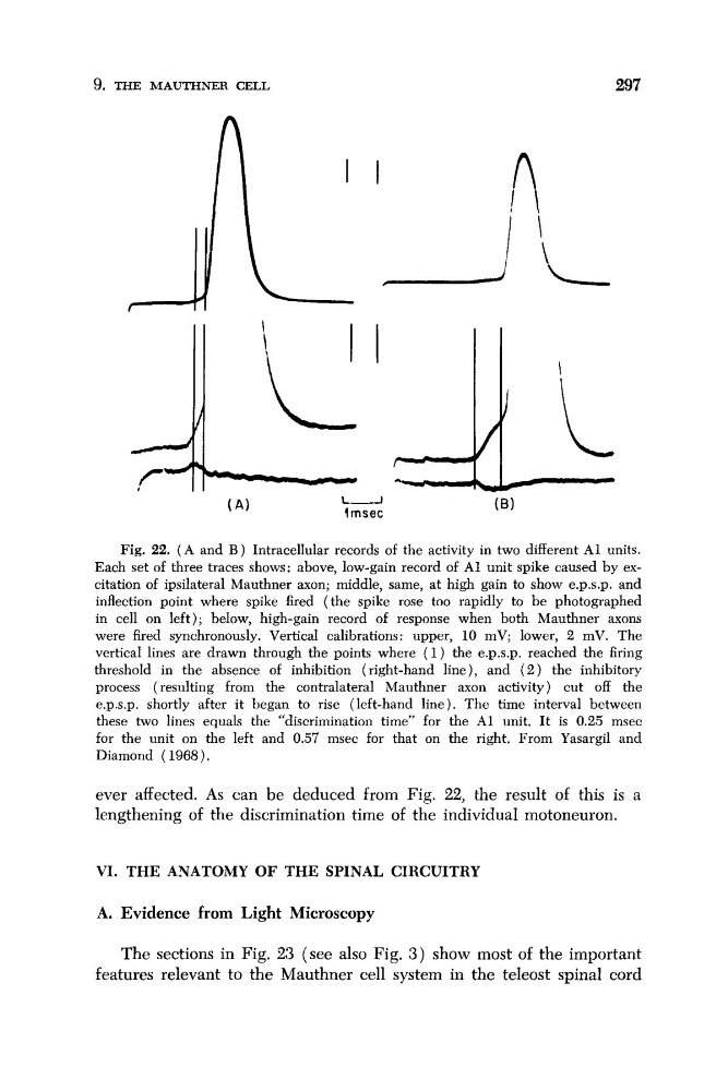
Fig. 22. ( A and B ) Intracellular records of the activity in two different A1 units.
Each set of three traces shows: above, low-gain record of A1 unit spike caused by excitation of ipsilateral Mauthner axon; middle, same, at high gain to show e.p.s.p. and
inflection point where spike fired (the spike rose too rapidly to be photographed
in cell on left); below, high-gain record of response when both Mauthner axons
were fired synchronously. Vertical calibrations: upper, 10 mV; lower, 2 mV. The
vertical lines are drawn through the points where ( 1 ) the e.p.s.p. reached the firing
threshold in the absence of inhibition (right-hand line), and ( 2 ) the inhibitory
process (resulting from the contralateral Mauthner axon activity) cut off the
e.p.s.p. shortly after it began to rise (left-hand line). The time interval between
these two lines equals the “discrimination time” for the A1 unit. It is 0.25 msec
for the unit on the left and 0.57 msec for that on the right, From Yasargil and
Diamond ( 1968).
ever affected. As can be deduced from Fig. 22, the result of this is a
lengthening of the discrimination time of the individual motoneuron.
VI. THE ANATOMY OF THE SPINAL CIRCUITRY
A. Evidence from Light Microscopy
The sections in Fig. 23 (see also Fig. 3 ) show most of the important
features relevant to the Mauthner cell system in the teleost spinal cord
297
U
(msec
Fig. 22. ( A and B ) Intracellular records of the activity in two different A1 units.
Each set of three traces shows: above, low-gain record of A1 unit spike caused by excitation of ipsilateral Mauthner axon; middle, same, at high gain to show e.p.s.p. and
inflection point where spike fired (the spike rose too rapidly to be photographed
in cell on left); below, high-gain record of response when both Mauthner axons
were fired synchronously. Vertical calibrations: upper, 10 mV; lower, 2 mV. The
vertical lines are drawn through the points where ( 1 ) the e.p.s.p. reached the firing
threshold in the absence of inhibition (right-hand line), and ( 2 ) the inhibitory
process (resulting from the contralateral Mauthner axon activity) cut off the
e.p.s.p. shortly after it began to rise (left-hand line). The time interval between
these two lines equals the “discrimination time” for the A1 unit. It is 0.25 msec
for the unit on the left and 0.57 msec for that on the right, From Yasargil and
Diamond ( 1968).
ever affected. As can be deduced from Fig. 22, the result of this is a
lengthening of the discrimination time of the individual motoneuron.
VI. THE ANATOMY OF THE SPINAL CIRCUITRY
A. Evidence from Light Microscopy
The sections in Fig. 23 (see also Fig. 3 ) show most of the important
features relevant to the Mauthner cell system in the teleost spinal cord