

246
AKE FLOCK
asymmetrically placed kinocilium ( Fig. 4). Neighboring cells have opposite orientation, a well-known fact from transmission microscopy, with
every other cell facing toward the tail or toward the head (Fig. 4).
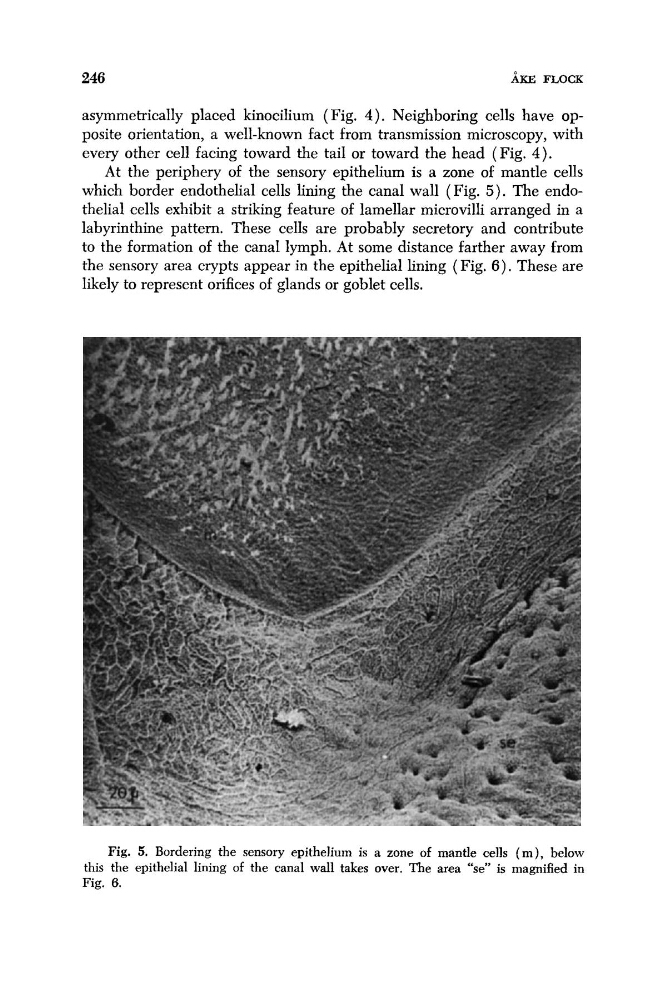
At the periphery of the sensory epithelium is a zone of mantle cells
which border endothelial cells lining the canal wall (Fig. 5 ) . The endothelial cells exhibit a striking feature of lamellar microvilli arranged in a
labyrinthine pattern. These cells are probably secretory and contribute
to the formation of the canal lymph. At some distance farther away from
the sensory area crypts appear in the epithelial lining ( Fig. 6 ) . These are
likely to represent orifices of glands or goblet cells.
Fig. 5. Bordering the sensory epithelium is a zone of mantle cells ( m ) , below
this the epithelial lining of the canal wall takes over. The area "se" is magnified in
Fig. 6.
AKE FLOCK
asymmetrically placed kinocilium ( Fig. 4). Neighboring cells have opposite orientation, a well-known fact from transmission microscopy, with
every other cell facing toward the tail or toward the head (Fig. 4).
At the periphery of the sensory epithelium is a zone of mantle cells
which border endothelial cells lining the canal wall (Fig. 5 ) . The endothelial cells exhibit a striking feature of lamellar microvilli arranged in a
labyrinthine pattern. These cells are probably secretory and contribute
to the formation of the canal lymph. At some distance farther away from
the sensory area crypts appear in the epithelial lining ( Fig. 6 ) . These are
likely to represent orifices of glands or goblet cells.
Fig. 5. Bordering the sensory epithelium is a zone of mantle cells ( m ) , below
this the epithelial lining of the canal wall takes over. The area "se" is magnified in
Fig. 6.