

222
0. LOWENSTEIN
Medial
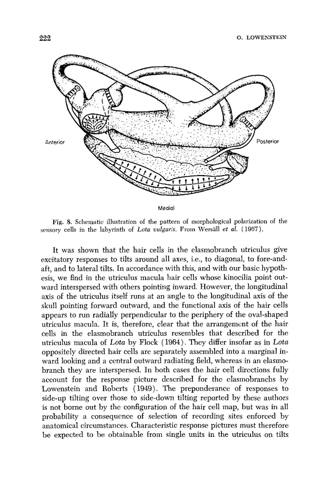
Fig. 8. Schematic illustration of the pattern of morphological polarization of the
sensory cells in the labyrinth of Lota uulgaris. From Wersall et al. (1967).
It was shown that the hair cells in the elasmobranch utriculus give
excitatory responses to tilts around all axes, i.e., to diagonal, to fore-andaft, and to lateral tilts. In accordance with this, and with our basic hypothesis, we find in the utriculus macula hair cells whose kinocilia point outward interspersed with others pointing inward. However, the longitudinal
axis of the utriculus itself runs at an angle to the longitudinal axis of the
skull pointing forward outward, and the functional axis of the hair cells
appears to run radially perpendicular to the periphery of the oval-shaped
utriculus macula. It is, therefore, clear that the arrangement of the hair
cells in the elasmobranch utriculus resembles that described for the
utriculus macula of Lota by Flock (1964). They differ insofar as in Lota
oppositely directed hair cells are separately assembled into a marginal inward looking and a central outward radiating field, whereas in an elasmobranch they are interspersed. In both cases the hair cell directions fully
account for the response picture described for the elasmobranchs by
Lowenstein and Roberts ( 1949). The preponderance of responses to
side-up tilting over those to side-down tilting reported by these authors
is not borne out by the configuration of the hair cell map, but was in all
probability a consequence of selection of recording sites enforced by
anatomical circumstances. Characteristic response pictures must therefore
be expected to be obtainable from single units in the utriculus on tilts
0. LOWENSTEIN
Medial
Fig. 8. Schematic illustration of the pattern of morphological polarization of the
sensory cells in the labyrinth of Lota uulgaris. From Wersall et al. (1967).
It was shown that the hair cells in the elasmobranch utriculus give
excitatory responses to tilts around all axes, i.e., to diagonal, to fore-andaft, and to lateral tilts. In accordance with this, and with our basic hypothesis, we find in the utriculus macula hair cells whose kinocilia point outward interspersed with others pointing inward. However, the longitudinal
axis of the utriculus itself runs at an angle to the longitudinal axis of the
skull pointing forward outward, and the functional axis of the hair cells
appears to run radially perpendicular to the periphery of the oval-shaped
utriculus macula. It is, therefore, clear that the arrangement of the hair
cells in the elasmobranch utriculus resembles that described for the
utriculus macula of Lota by Flock (1964). They differ insofar as in Lota
oppositely directed hair cells are separately assembled into a marginal inward looking and a central outward radiating field, whereas in an elasmobranch they are interspersed. In both cases the hair cell directions fully
account for the response picture described for the elasmobranchs by
Lowenstein and Roberts ( 1949). The preponderance of responses to
side-up tilting over those to side-down tilting reported by these authors
is not borne out by the configuration of the hair cell map, but was in all
probability a consequence of selection of recording sites enforced by
anatomical circumstances. Characteristic response pictures must therefore
be expected to be obtainable from single units in the utriculus on tilts