

7. THE LABYRINTH
213
tudinal fibrillae. Their number varies between a few tens and over a
hundred and their length is usuaily graded, their tips forming a slope
away from the single so-called kinocilium. This kinocilium is rooted in
the cytoplasm outside the cuticular plate by means of a typical basal
body or centriole. The kinocilium is, in fact, a true cilium, characterized
by nine peripheral and two central double longitudinal filaments. The
kinocilium is often much longer than the longest stereocilia. The centriole is composed of nine triplet tubules in continuity with the double
filaments of the ciliary shaft. On the side facing away from the cuticular
plate and from the stereocilia, one of the triplet tubules has a lateral excrescense of electron-dense material, the so-called basal foot. The arrangement of hair processes is thus strictly polarized, and it has been shown
that this polarization is of fundamental functional significance ( Lowenstein et al., 1964).
The hair cells of the cristae and maculae have been found to be
oriented within the sensory epithelia in a constant characteristic pattern.
It is assumed that the mechanical deformation of the hair bundle, or at
least the shearing of the kinocilial base in the direction of the basal foot,
has an excitatory effect and deformation in the opposite direction an inhibitory effect on the activity of the hair cell and its associated sensory
neuron.
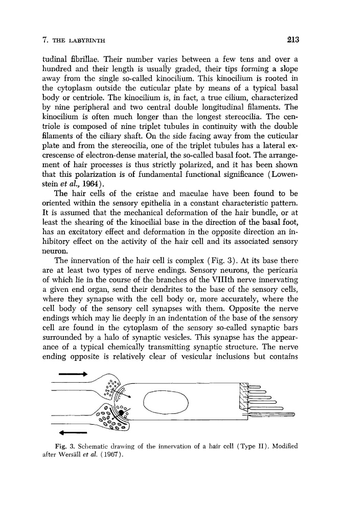
The innervation of the hair cell is complex (Fig. 3 ) . At its base there
are at least two types of nerve endings. Sensory neurons, the pericaria
of which lie in the course of the branches of the VIIIth nerve innervating
a given end organ, send their dendrites to the base of the sensory cells,
where they synapse with the cell body or, more accurately, where the
cell body of the sensory cell synapses with them. Opposite the nerve
endings which may lie deeply in an indentation of the base of the sensory
cell are found in the cytoplasm of the sensory so-called synaptic bars
surrounded by a halo of synaptic vesicles. This synapse has the appearance of a typical chemically transmitting synaptic structure. The nerve
ending opposite is relatively clear of vesicular inclusions but contains
_Fig. 3. Schematic drawing of the innervation of a hair cell (Type 11). Modified
after Wersall et al. ( 1967).
213
tudinal fibrillae. Their number varies between a few tens and over a
hundred and their length is usuaily graded, their tips forming a slope
away from the single so-called kinocilium. This kinocilium is rooted in
the cytoplasm outside the cuticular plate by means of a typical basal
body or centriole. The kinocilium is, in fact, a true cilium, characterized
by nine peripheral and two central double longitudinal filaments. The
kinocilium is often much longer than the longest stereocilia. The centriole is composed of nine triplet tubules in continuity with the double
filaments of the ciliary shaft. On the side facing away from the cuticular
plate and from the stereocilia, one of the triplet tubules has a lateral excrescense of electron-dense material, the so-called basal foot. The arrangement of hair processes is thus strictly polarized, and it has been shown
that this polarization is of fundamental functional significance ( Lowenstein et al., 1964).
The hair cells of the cristae and maculae have been found to be
oriented within the sensory epithelia in a constant characteristic pattern.
It is assumed that the mechanical deformation of the hair bundle, or at
least the shearing of the kinocilial base in the direction of the basal foot,
has an excitatory effect and deformation in the opposite direction an inhibitory effect on the activity of the hair cell and its associated sensory
neuron.
The innervation of the hair cell is complex (Fig. 3 ) . At its base there
are at least two types of nerve endings. Sensory neurons, the pericaria
of which lie in the course of the branches of the VIIIth nerve innervating
a given end organ, send their dendrites to the base of the sensory cells,
where they synapse with the cell body or, more accurately, where the
cell body of the sensory cell synapses with them. Opposite the nerve
endings which may lie deeply in an indentation of the base of the sensory
cell are found in the cytoplasm of the sensory so-called synaptic bars
surrounded by a halo of synaptic vesicles. This synapse has the appearance of a typical chemically transmitting synaptic structure. The nerve
ending opposite is relatively clear of vesicular inclusions but contains
_Fig. 3. Schematic drawing of the innervation of a hair cell (Type 11). Modified
after Wersall et al. ( 1967).