

6. SOUND PRODUCTION AND DETECTION
165
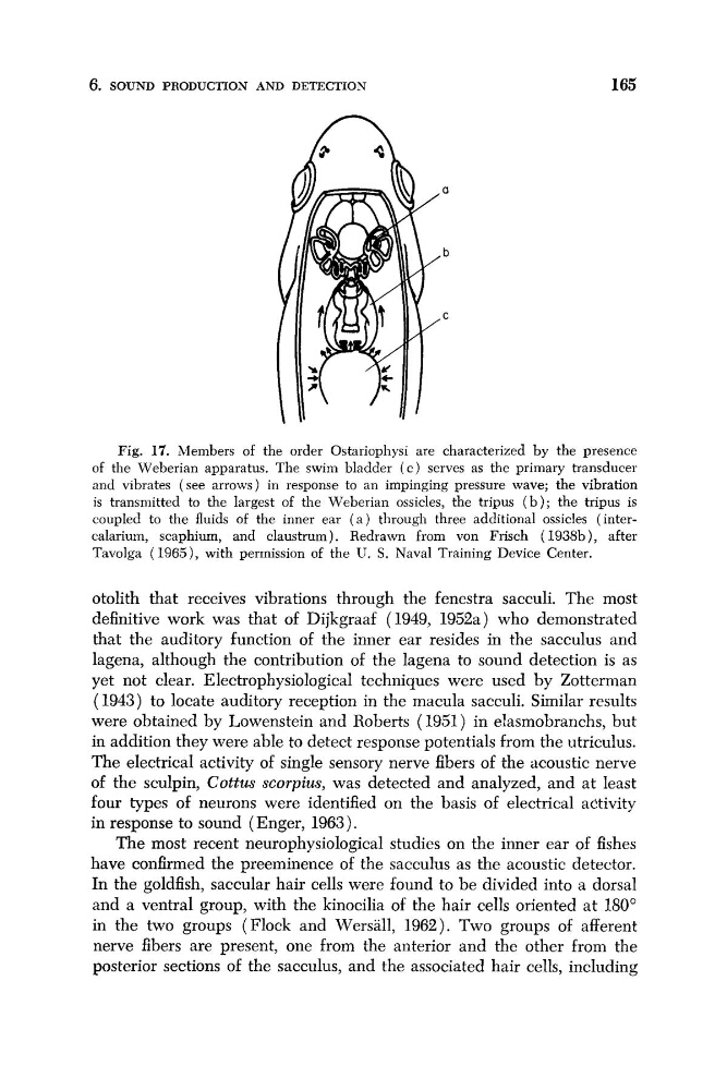
Fig. 17. Members of the order Ostariophysi are characterized by the presence
of the Weberian apparatus. The swim bladder ( c ) serves as the primary transducer
and vibrates (see arrows) in response to an impinging pressure wave; the vibration
is transmitted to the largest of the Weberian ossicles, the tripus ( b ) ; the tripus is
coupled to the fluids of the inner ear ( a ) through three additional ossicles (intercalariurn, scaphium, and claustrum). Redrawn from von Frisch (1938b), after
Tavolga (1965), with permission of the U. S. Naval Training Device Center.
otolith that receives vibrations through the fenestra sacculi. The most
definitive work was that of Dijkgraaf (1949, 1952a) who demonstrated
that the auditory function of the inner ear resides in the sacculus and
lagena, although the contribution of the lagena to sound detection is as
yet not clear. Electrophysiological techniques were used by Zotterman
(1943) to locate auditory reception in the macula sacculi. Similar results
were obtained by Lowenstein and Roberts (1951) in elasmobranchs, but
in addition they were able to detect response potentials from the utriculus.
The electrical activity of single sensory nerve fibers of the acoustic nerve
of the sculpin, Cottus scorpius, was detected and analyzed, and at least
four types of neurons were identified on the basis of electrical activity
in response to sound (Enger, 1963).
The most recent neurophysiological studies on the inner ear of fishes
have confirmed the preeminence of the sacculus as the acoustic detector.
In the goldfish, saccular hair cells were found to be divided into a dorsal
and a ventral group, with the kinocilia of the hair cells oriented at 180"
in the two groups (Flock and Wersall, 1962). Two groups of afferent
nerve fibers are present, one from the anterior and the other from the
posterior sections of the sacculus, and the associated hair cells, including
165
Fig. 17. Members of the order Ostariophysi are characterized by the presence
of the Weberian apparatus. The swim bladder ( c ) serves as the primary transducer
and vibrates (see arrows) in response to an impinging pressure wave; the vibration
is transmitted to the largest of the Weberian ossicles, the tripus ( b ) ; the tripus is
coupled to the fluids of the inner ear ( a ) through three additional ossicles (intercalariurn, scaphium, and claustrum). Redrawn from von Frisch (1938b), after
Tavolga (1965), with permission of the U. S. Naval Training Device Center.
otolith that receives vibrations through the fenestra sacculi. The most
definitive work was that of Dijkgraaf (1949, 1952a) who demonstrated
that the auditory function of the inner ear resides in the sacculus and
lagena, although the contribution of the lagena to sound detection is as
yet not clear. Electrophysiological techniques were used by Zotterman
(1943) to locate auditory reception in the macula sacculi. Similar results
were obtained by Lowenstein and Roberts (1951) in elasmobranchs, but
in addition they were able to detect response potentials from the utriculus.
The electrical activity of single sensory nerve fibers of the acoustic nerve
of the sculpin, Cottus scorpius, was detected and analyzed, and at least
four types of neurons were identified on the basis of electrical activity
in response to sound (Enger, 1963).
The most recent neurophysiological studies on the inner ear of fishes
have confirmed the preeminence of the sacculus as the acoustic detector.
In the goldfish, saccular hair cells were found to be divided into a dorsal
and a ventral group, with the kinocilia of the hair cells oriented at 180"
in the two groups (Flock and Wersall, 1962). Two groups of afferent
nerve fibers are present, one from the anterior and the other from the
posterior sections of the sacculus, and the associated hair cells, including