

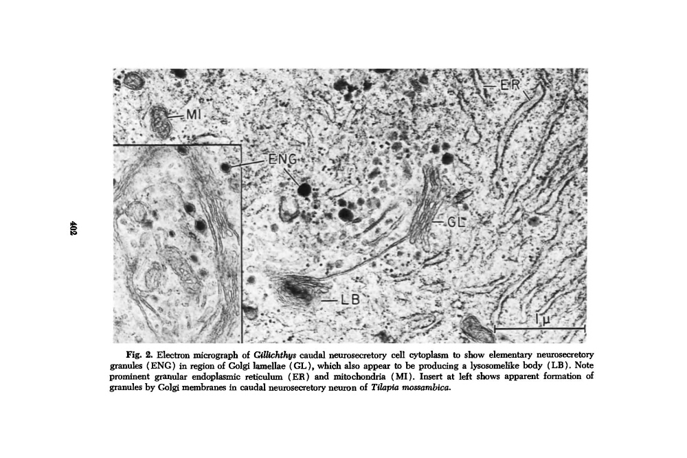
Fig. 2. Electron micrograph of GiUichthvs caudal neurosecretory cell cytoplasm to show elementary neurosecretory
granules (ENG) in region of Golgi lamellae ( GL), which also appear to be producing a lysosomelike body (LB). Note
prominent granular endoplasmic reticulum (ER) and mitochondria (MI). Insert at left shows apparent formation of
granules by Golgi membranes in caudal neurosecretory neuron of Tilnpia mossambicu.
granules (ENG) in region of Golgi lamellae ( GL), which also appear to be producing a lysosomelike body (LB). Note
prominent granular endoplasmic reticulum (ER) and mitochondria (MI). Insert at left shows apparent formation of
granules by Golgi membranes in caudal neurosecretory neuron of Tilnpia mossambicu.