

8. UROPHYSIS AND CAUDAL NEUROSECRETORY SYSTEM
401
the most useful systematic comparative anatomic treatments are those of
Fridberg (1963) and McLean ( 1965).
In this chapter we shall attempt to survey the information currently
available on the caudal neurosecretory system and to raise issues and
questions which may aid further study directed toward the definitive
functional elucidation of this fascinating neuroendocrine apparatus.
11. ANATOMY AND OCCURRENCE OF THE CAUDAL
NEUROSECRETORY SYSTEM
A. Caudal Neurosecretory System in Teleosts and Elasmobranchs
1. TELEOSTS
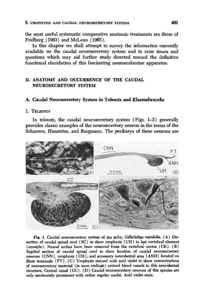
In teleosts, the caudal neurosecretory system (Figs. 1-3) generally
provides classic examples of the neurosecretory neuron in the terms of the
Scharrers, Hanstrom, and Bargmann. The perikarya of these neurons are
Fig. 1. Caudal neurosecretory system of the goby, Gillichthys mirabi2is. (A) Dissection of caudal spinal cord (SC) to show urophysis ( U H ) in last vertebral element
(urostyle). Neural arches have been removed from the vertebral centra (CE ). (B)
Sagittal section of caudal spinal cord to show location of caudal neurosecretory
neurons (CNN), urophysis ( UH), and accessory neurohemal area (ANH) located on
mum terminale (FT). ( C ) Urophysis stained with acid violet to show concentrations
of neurosecretory material (in axon endings) around blood vessels in this neurohemal
structure. Central canal ( CC) . ( D ) Caudal neurosecretory neurons of this species are
only moderately prominent with rather regular nuclei. Acid violet stain.
401
the most useful systematic comparative anatomic treatments are those of
Fridberg (1963) and McLean ( 1965).
In this chapter we shall attempt to survey the information currently
available on the caudal neurosecretory system and to raise issues and
questions which may aid further study directed toward the definitive
functional elucidation of this fascinating neuroendocrine apparatus.
11. ANATOMY AND OCCURRENCE OF THE CAUDAL
NEUROSECRETORY SYSTEM
A. Caudal Neurosecretory System in Teleosts and Elasmobranchs
1. TELEOSTS
In teleosts, the caudal neurosecretory system (Figs. 1-3) generally
provides classic examples of the neurosecretory neuron in the terms of the
Scharrers, Hanstrom, and Bargmann. The perikarya of these neurons are
Fig. 1. Caudal neurosecretory system of the goby, Gillichthys mirabi2is. (A) Dissection of caudal spinal cord (SC) to show urophysis ( U H ) in last vertebral element
(urostyle). Neural arches have been removed from the vertebral centra (CE ). (B)
Sagittal section of caudal spinal cord to show location of caudal neurosecretory
neurons (CNN), urophysis ( UH), and accessory neurohemal area (ANH) located on
mum terminale (FT). ( C ) Urophysis stained with acid violet to show concentrations
of neurosecretory material (in axon endings) around blood vessels in this neurohemal
structure. Central canal ( CC) . ( D ) Caudal neurosecretory neurons of this species are
only moderately prominent with rather regular nuclei. Acid violet stain.