

384
D. HAROLD COPP
basibranchial and ceratobranchial cartilages and the coracobranchial
muscle. The gland is highly vascularized and consists of many follicles
such as those shown in Fig. 2. These are about 125 p in diameter and lined
with columnar epithelium. There is granular material i n the follicular
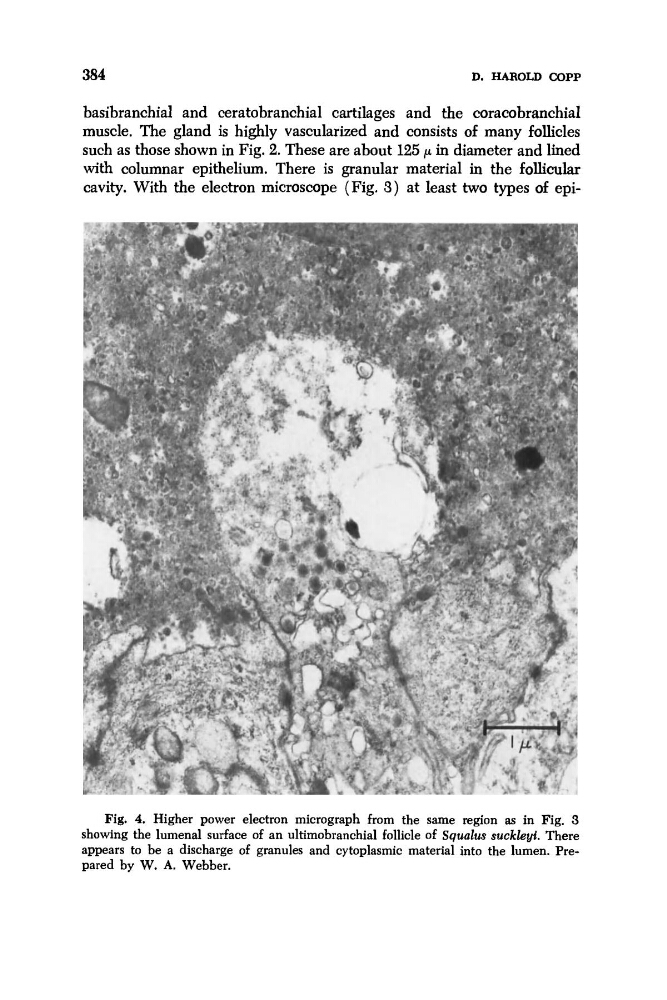
cavity. With the electron microscope (Fig. 3 ) at least two types of epiFig. 4. Higher power electron micrograph from the same region as in Fig. 3
showing the lumenal surface of an ultimobranchial follicle of Squalzrs suckleyi. There
appears to be a discharge of granules and cytoplasmic material into the lumen. Prepared by W. A. Webber.
D. HAROLD COPP
basibranchial and ceratobranchial cartilages and the coracobranchial
muscle. The gland is highly vascularized and consists of many follicles
such as those shown in Fig. 2. These are about 125 p in diameter and lined
with columnar epithelium. There is granular material i n the follicular
cavity. With the electron microscope (Fig. 3 ) at least two types of epiFig. 4. Higher power electron micrograph from the same region as in Fig. 3
showing the lumenal surface of an ultimobranchial follicle of Squalzrs suckleyi. There
appears to be a discharge of granules and cytoplasmic material into the lumen. Prepared by W. A. Webber.