

8. THE REGULATION OF BREATHING
313
Interarcualis
Lev palatoquadrati
Lev hyomandibulae
Subspinalis
dorsalis ."
Pre-orbitalis /
L J
Y I L .
Coraco-mandibularis
CoracoCoraco-bkchiales COmmuniS
hyoideus
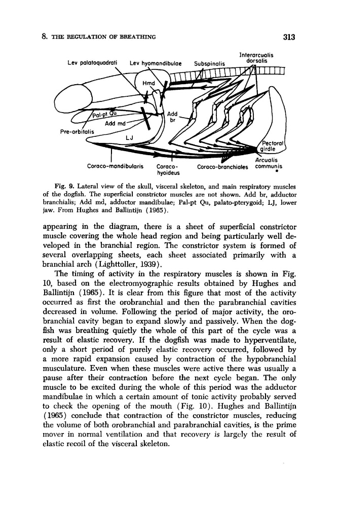
Fig. 9. Lateral view of the skull, visceral skeleton, and main respiratory muscles
of the dogfish. The superficial constrictor muscles are not shown. Add br, adductor
branchialis; Add md, adductor mandibulae; Pal-pt Qu, palato-pterygoid; LJ, lower
jaw. From Hughes and Ballintijn ( 1965).
appearing in the diagram, there is a sheet of superficial constrictor
muscle covering the whole head region and being particularly well developed in the branchial region. The constrictor system is formed of
several overlapping sheets, each sheet associated primarily with a
branchial arch ( Lighttoller, 1939).
The timing of activity in the respiratory muscles is shown in Fig.
10, based on the electromyographic results obtained by Hughes and
Ballintijn (1965). It is clear from this figure that most of the activity
occurred as first the orobranchial and then the parabranchial cavities
decreased in volume. Following the period of major activity, the orobranchial cavity began to expand slowly and passively. When the dogfish was breathing quietly the whole of this part of the cycle was a
result of elastic recovery. If the dogfish was made to hyperventilate,
only a short period of purely elastic recovery occurred, followed by
a more rapid expansion caused by contraction of the hypobranchial
musculature. Even when these muscles were active there was usually a
pause after their contraction before the next cycle began. The only
muscle to be excited during the whole of this period was the adductor
mandibulae in which a certain amount of tonic activity probably sewed
to check the opening of the mouth (Fig. 10). Hughes and Ballintijn
(1965) conclude that contraction of the constrictor muscles, reducing
the volume of both orobranchial and parabranchial cavities, is the prime
mover in normal ventilation and that recovery is largely the result of
elastic recoil of the visceral skeleton.
313
Interarcualis
Lev palatoquadrati
Lev hyomandibulae
Subspinalis
dorsalis ."
Pre-orbitalis /
L J
Y I L .
Coraco-mandibularis
CoracoCoraco-bkchiales COmmuniS
hyoideus
Fig. 9. Lateral view of the skull, visceral skeleton, and main respiratory muscles
of the dogfish. The superficial constrictor muscles are not shown. Add br, adductor
branchialis; Add md, adductor mandibulae; Pal-pt Qu, palato-pterygoid; LJ, lower
jaw. From Hughes and Ballintijn ( 1965).
appearing in the diagram, there is a sheet of superficial constrictor
muscle covering the whole head region and being particularly well developed in the branchial region. The constrictor system is formed of
several overlapping sheets, each sheet associated primarily with a
branchial arch ( Lighttoller, 1939).
The timing of activity in the respiratory muscles is shown in Fig.
10, based on the electromyographic results obtained by Hughes and
Ballintijn (1965). It is clear from this figure that most of the activity
occurred as first the orobranchial and then the parabranchial cavities
decreased in volume. Following the period of major activity, the orobranchial cavity began to expand slowly and passively. When the dogfish was breathing quietly the whole of this part of the cycle was a
result of elastic recovery. If the dogfish was made to hyperventilate,
only a short period of purely elastic recovery occurred, followed by
a more rapid expansion caused by contraction of the hypobranchial
musculature. Even when these muscles were active there was usually a
pause after their contraction before the next cycle began. The only
muscle to be excited during the whole of this period was the adductor
mandibulae in which a certain amount of tonic activity probably sewed
to check the opening of the mouth (Fig. 10). Hughes and Ballintijn
(1965) conclude that contraction of the constrictor muscles, reducing
the volume of both orobranchial and parabranchial cavities, is the prime
mover in normal ventilation and that recovery is largely the result of
elastic recoil of the visceral skeleton.