

6. PROPERTIES OF FISH HEMOGLOBINS
211
formation of horse hemoglobin chains (Perutz et al., 1968), sperm whale
myoglobin (Kendrew et aZ., 1960), the hemoglobins of the marine annelid, Glycera (Padlan and Love, 1968), and that of the insect Chirommus (Huber et al., 1968) lends confidence to the belief that all hemoglobin polypeptide chains, both vertebrate and invertebrate, are arranged
similarly and all are derived from a common ancestral form. Therefore,
we have every expectation that X-ray diffraction and amino acid sequence studies will show fish hemoglobins to possess many features in
common with the better studied mammalian pigments. Nevertheless,
this should not obscure the fact that fish hemoglobins possess many
unique features, both in their physiology and their primary structure.
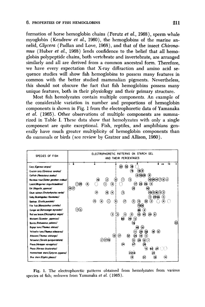
Most fish hemolyzates contain multiple components. An example of
the considerable variation in number and proportions of hemoglobin
components is shown in Fig. 1 from the electrophoretic data of Yamanaka
et al. (1965). Other observations of multiple components are summarized in Table I. These data show that hemolyzates with only a single
component are quite exceptional. Fish, reptiles, and amphibians generally have much greater multiplicity of hemoglobin components than
do mammals or birds (see review by Gratzer and Allison, 1960).
ELECTROPHORETIC PATTERNS ON STARCH GEL
AND THEIR PERCENTAGES
Fig. 1. The electrophoretic patterns obtained from hemolyzates from various
species of fish; redrawn from Yamanaka et al. (1965).
211
formation of horse hemoglobin chains (Perutz et al., 1968), sperm whale
myoglobin (Kendrew et aZ., 1960), the hemoglobins of the marine annelid, Glycera (Padlan and Love, 1968), and that of the insect Chirommus (Huber et al., 1968) lends confidence to the belief that all hemoglobin polypeptide chains, both vertebrate and invertebrate, are arranged
similarly and all are derived from a common ancestral form. Therefore,
we have every expectation that X-ray diffraction and amino acid sequence studies will show fish hemoglobins to possess many features in
common with the better studied mammalian pigments. Nevertheless,
this should not obscure the fact that fish hemoglobins possess many
unique features, both in their physiology and their primary structure.
Most fish hemolyzates contain multiple components. An example of
the considerable variation in number and proportions of hemoglobin
components is shown in Fig. 1 from the electrophoretic data of Yamanaka
et al. (1965). Other observations of multiple components are summarized in Table I. These data show that hemolyzates with only a single
component are quite exceptional. Fish, reptiles, and amphibians generally have much greater multiplicity of hemoglobin components than
do mammals or birds (see review by Gratzer and Allison, 1960).
ELECTROPHORETIC PATTERNS ON STARCH GEL
AND THEIR PERCENTAGES
Fig. 1. The electrophoretic patterns obtained from hemolyzates from various
species of fish; redrawn from Yamanaka et al. (1965).