

146
D. J. RANDALL
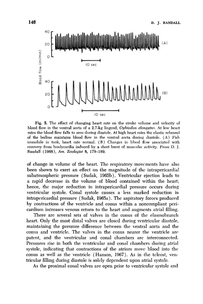
Fig. 5. The effect of changing heart rate on the stroke volume and velocity of
blood flow in the ventral aorta of a 2.7-kg lingcod, Ophiodon eZongatrcP. At low heart
rates the blood flow falls to zero during diastole. At high heart rates the elastic rebound
of the bulbus maintains blood flow in the ventral aorta during diastole. ( A ) Fish
immobile in tank, heart rate normal. ( B ) Changes in blood flow associated with
recovery from bradycardia induced by a short burst of muscnlar activity. From D. J.
Randall (1968), Am. Zoologist 8, 179-189.
of change in volume of the heart. The respiratory movcments have also
been shown to exert an effect on the magnitude of the intrapericardial
subatmospheric pressure ( Sudak, 1965b). Ventricular ejection leads to
a rapid decrease in the volume of blood contained within the heart;
hence, the major reduction in intrapericardial pressure occurs during
ventricular systole. Conal systole causes a less marked reduction in
intrapericardial pressure ( Sudak, 1965a ) . Thc aspiratory forces produced
by contractions of the ventricle and conus within a noncompliant pericardium increases venous return to the heart and augments atrial filling.
There are several sets of valves in the conus of the elasmobranch
heart. Only the most distal valves are closcd during ventricular diastole,
maintaining the pressure difference between the ventral aorta and the
conus and ventricle. The valves in the conus nearer the ventricle are
patent, and the ventricular and conal chambers arc interconnected.
Pressures rise in both the ventricular and conal chambers during atrial
systole, indicating that contractions of the atrium move blood into the
conus as well as the ventricle (Hanson, 1967). As in the tcleost, ventricular filling during diastole is solely dependent upon atrial systole.
As the proximal conal valves are open prior to ventricular systole and
D. J. RANDALL
Fig. 5. The effect of changing heart rate on the stroke volume and velocity of
blood flow in the ventral aorta of a 2.7-kg lingcod, Ophiodon eZongatrcP. At low heart
rates the blood flow falls to zero during diastole. At high heart rates the elastic rebound
of the bulbus maintains blood flow in the ventral aorta during diastole. ( A ) Fish
immobile in tank, heart rate normal. ( B ) Changes in blood flow associated with
recovery from bradycardia induced by a short burst of muscnlar activity. From D. J.
Randall (1968), Am. Zoologist 8, 179-189.
of change in volume of the heart. The respiratory movcments have also
been shown to exert an effect on the magnitude of the intrapericardial
subatmospheric pressure ( Sudak, 1965b). Ventricular ejection leads to
a rapid decrease in the volume of blood contained within the heart;
hence, the major reduction in intrapericardial pressure occurs during
ventricular systole. Conal systole causes a less marked reduction in
intrapericardial pressure ( Sudak, 1965a ) . Thc aspiratory forces produced
by contractions of the ventricle and conus within a noncompliant pericardium increases venous return to the heart and augments atrial filling.
There are several sets of valves in the conus of the elasmobranch
heart. Only the most distal valves are closcd during ventricular diastole,
maintaining the pressure difference between the ventral aorta and the
conus and ventricle. The valves in the conus nearer the ventricle are
patent, and the ventricular and conal chambers arc interconnected.
Pressures rise in both the ventricular and conal chambers during atrial
systole, indicating that contractions of the atrium move blood into the
conus as well as the ventricle (Hanson, 1967). As in the tcleost, ventricular filling during diastole is solely dependent upon atrial systole.
As the proximal conal valves are open prior to ventricular systole and