

144
D. J. RANDALL
Vent r it le
0
I sec
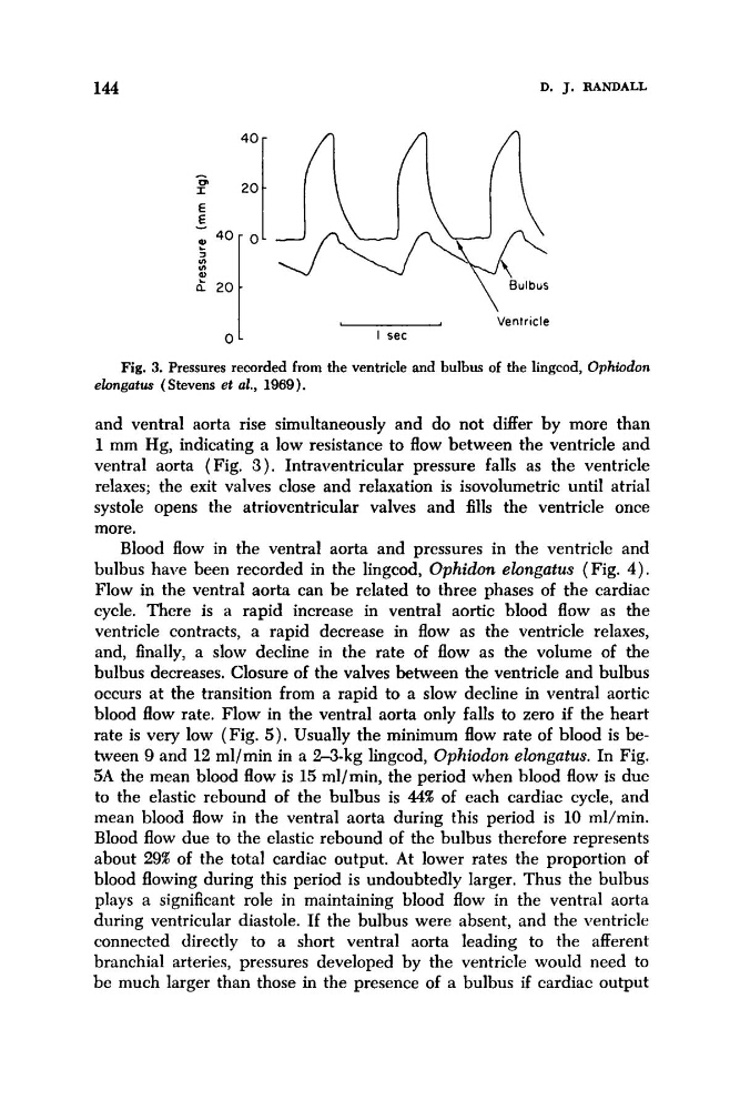
Fig. 3. Pressures recorded from the ventricle and bulbus of the lingcod, Ophiodon
elongatus (Stevens et al., 1969).
and ventral aorta rise simultaneously and do not differ by more than
1 mm Hg, indicating a low resistance to flow between the ventricle and
ventral aorta (Fig. 3). Intraventricular pressure falls as the ventricle
relaxes; the exit valves close and relaxation is isovolumetric until atrial
systole opens the atrioventricular valves and fills the ventricle once
more.
Blood flow in the ventral aorta and pressures in the ventricle and
bulbus have been recorded in the lingcod, Ophidon ebngatus (Fig. 4).
Flow in the ventral aorta can be related to three phases of the cardiac
cycle. There is a rapid increase in ventral aortic blood flow as the
ventricle contracts, a rapid decrease in flow as the ventricle relaxes,
and, finally, a slow decline in the rate of flow as the volume of the
bulbus decreases. Closure of the valves between the ventricle and bulbus
occurs at the transition from a rapid to a slow decline in ventral aortic
blood flow rate. Flow in the ventral aorta only falls to zero if the heart
rate is very low (Fig. 5). Usually the minimum flow rate of blood is between 9 and 12 ml/min in a 2-3-kg lingcod, Ophiodon ebngatus. In Fig.
5A the mean blood flow is 15 ml/min, the period when blood flow is due
to the elastic rebound of the bulbus is 44% of each cardiac cycle, and
mean blood flow in the ventral aorta during this period is 10 ml/min.
Blood flow due to the elastic rebound of the bulbus therefore represents
about 291% of the total cardiac output. At lower rates the proportion of
blood flowing during this period is undoubtedly larger. Thus the bulbus
plays a significant role in maintaining blood flow in the ventral aorta
during ventricular diastole. If the bulbus were absent, and the ventricle
connected directly to a short ventral aorta leading to the afferent
branchial arteries, pressures developed by the ventricle would need to
be much larger than those in the presence of a bulbus if cardiac output
D. J. RANDALL
Vent r it le
0
I sec
Fig. 3. Pressures recorded from the ventricle and bulbus of the lingcod, Ophiodon
elongatus (Stevens et al., 1969).
and ventral aorta rise simultaneously and do not differ by more than
1 mm Hg, indicating a low resistance to flow between the ventricle and
ventral aorta (Fig. 3). Intraventricular pressure falls as the ventricle
relaxes; the exit valves close and relaxation is isovolumetric until atrial
systole opens the atrioventricular valves and fills the ventricle once
more.
Blood flow in the ventral aorta and pressures in the ventricle and
bulbus have been recorded in the lingcod, Ophidon ebngatus (Fig. 4).
Flow in the ventral aorta can be related to three phases of the cardiac
cycle. There is a rapid increase in ventral aortic blood flow as the
ventricle contracts, a rapid decrease in flow as the ventricle relaxes,
and, finally, a slow decline in the rate of flow as the volume of the
bulbus decreases. Closure of the valves between the ventricle and bulbus
occurs at the transition from a rapid to a slow decline in ventral aortic
blood flow rate. Flow in the ventral aorta only falls to zero if the heart
rate is very low (Fig. 5). Usually the minimum flow rate of blood is between 9 and 12 ml/min in a 2-3-kg lingcod, Ophiodon ebngatus. In Fig.
5A the mean blood flow is 15 ml/min, the period when blood flow is due
to the elastic rebound of the bulbus is 44% of each cardiac cycle, and
mean blood flow in the ventral aorta during this period is 10 ml/min.
Blood flow due to the elastic rebound of the bulbus therefore represents
about 291% of the total cardiac output. At lower rates the proportion of
blood flowing during this period is undoubtedly larger. Thus the bulbus
plays a significant role in maintaining blood flow in the ventral aorta
during ventricular diastole. If the bulbus were absent, and the ventricle
connected directly to a short ventral aorta leading to the afferent
branchial arteries, pressures developed by the ventricle would need to
be much larger than those in the presence of a bulbus if cardiac output