

ihnd Thorpc, I !)(is ; Sticiventu.t8, 1970). Since Berrill's summary of the
Literature thore have been coiuiderablo advances in our knowledge of
the composition and formation of the test in various species which has
been enhanced by the us(? of txacer techniques and electron-microscopy.
AR the structure aud proccuses involved appear to be different in
different species, some of these are individually summarized below.
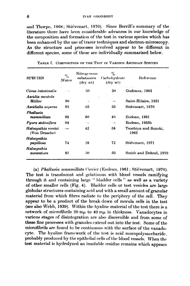
'rAnLE 1. C'OYPOWITION OP TIKR TlWl' I N VARIOUS ASCIDIAN SPECIES
Nitrogt W ) I I H
' 2,
'% ~rthatciiiccw ( ' ~ i i , l i ~ ) l i ~ ~ ~ ~ ~ i i , [ ,
1Ccfcwnco
SVIttW
8 PE(" ES
(dry wf 1
((try w( 1
Cwna in,testinnliR
50
50
Godoaux, 1963
Ascdia inerttitla
Aeeidiella avperuo
!)5
ti5
35
StiBvonart, 1970
Phollwia
mammi~lah
95
60
40
Endean, 1961
Miilloi~
!)O
-
Saint-Hilaire, 1031
Pyura atoloiiifera
!)4
-
-
E~ldert~l, 1'35513
HalocyntWiu. rorctzi
-
42
58
'rsuchiya and Suziiki,
Halocynthia
Halocynthin
(Von DraasclieI
1962
po pilloea
71
'
8
72
YtiBventtrt, 1'371
aurantiurn
81
50
50
Smith and Dehncl, 1970
(a) Phnlluttia r/tQnzrnillrxta ( 'uvivr ( 141t(let~ti, 1961 ; Htihwnart, 1970).
The test is translucent ant1 g ~ l i ~ t i ~ i ~ ~ ~
with blood V ~ ~ S C ~ S
ramifying
through it and containing large " bltdder d s " as well a8 a variety
of other sinaller cclls (Fig. 4). Ulatldcr cclls or test vcsiclcs are large
globular structures containing acid and with a small amount of granular
material from which fibrw radiate to the periphery of the cell. They
appear to be a product of the break-down of morula cells in the test
(see also Webb, 1939). Within the hyaline material of the test there is a
network of microfibrils 20 nip to 40 nip in thickness. Vanadocytew in
various stages of disint,egrat]ion are also discernible and from some of
these fine processes with granules cxtetd out into the test. Some of the
microfibrils Itre found to be continuous with t hc surface of the vanadocyte. Ttic hydine frantcbwork of the test is acid niric~p~lysaccharide.
probably produced by t h e epithc~lid cells of the blood vessels. When t h r
test material is hydrolyzed an insolublc reuidue remains which appears
Literature thore have been coiuiderablo advances in our knowledge of
the composition and formation of the test in various species which has
been enhanced by the us(? of txacer techniques and electron-microscopy.
AR the structure aud proccuses involved appear to be different in
different species, some of these are individually summarized below.
'rAnLE 1. C'OYPOWITION OP TIKR TlWl' I N VARIOUS ASCIDIAN SPECIES
Nitrogt W ) I I H
' 2,
'% ~rthatciiiccw ( ' ~ i i , l i ~ ) l i ~ ~ ~ ~ ~ i i , [ ,
1Ccfcwnco
SVIttW
8 PE(" ES
(dry wf 1
((try w( 1
Cwna in,testinnliR
50
50
Godoaux, 1963
Ascdia inerttitla
Aeeidiella avperuo
!)5
ti5
35
StiBvonart, 1970
Phollwia
mammi~lah
95
60
40
Endean, 1961
Miilloi~
!)O
-
Saint-Hilaire, 1031
Pyura atoloiiifera
!)4
-
-
E~ldert~l, 1'35513
HalocyntWiu. rorctzi
-
42
58
'rsuchiya and Suziiki,
Halocynthia
Halocynthin
(Von DraasclieI
1962
po pilloea
71
'
8
72
YtiBventtrt, 1'371
aurantiurn
81
50
50
Smith and Dehncl, 1970
(a) Phnlluttia r/tQnzrnillrxta ( 'uvivr ( 141t(let~ti, 1961 ; Htihwnart, 1970).
The test is translucent ant1 g ~ l i ~ t i ~ i ~ ~ ~
with blood V ~ ~ S C ~ S
ramifying
through it and containing large " bltdder d s " as well a8 a variety
of other sinaller cclls (Fig. 4). Ulatldcr cclls or test vcsiclcs are large
globular structures containing acid and with a small amount of granular
material from which fibrw radiate to the periphery of the cell. They
appear to be a product of the break-down of morula cells in the test
(see also Webb, 1939). Within the hyaline material of the test there is a
network of microfibrils 20 nip to 40 nip in thickness. Vanadocytew in
various stages of disint,egrat]ion are also discernible and from some of
these fine processes with granules cxtetd out into the test. Some of the
microfibrils Itre found to be continuous with t hc surface of the vanadocyte. Ttic hydine frantcbwork of the test is acid niric~p~lysaccharide.
probably produced by t h e epithc~lid cells of the blood vessels. When t h r
test material is hydrolyzed an insolublc reuidue remains which appears