

B
R
1
C
A
O1
4
d2
B
%
C
MG
9
1 0
F
F
- ss
0 5 m m
AA
OD R
I'
17
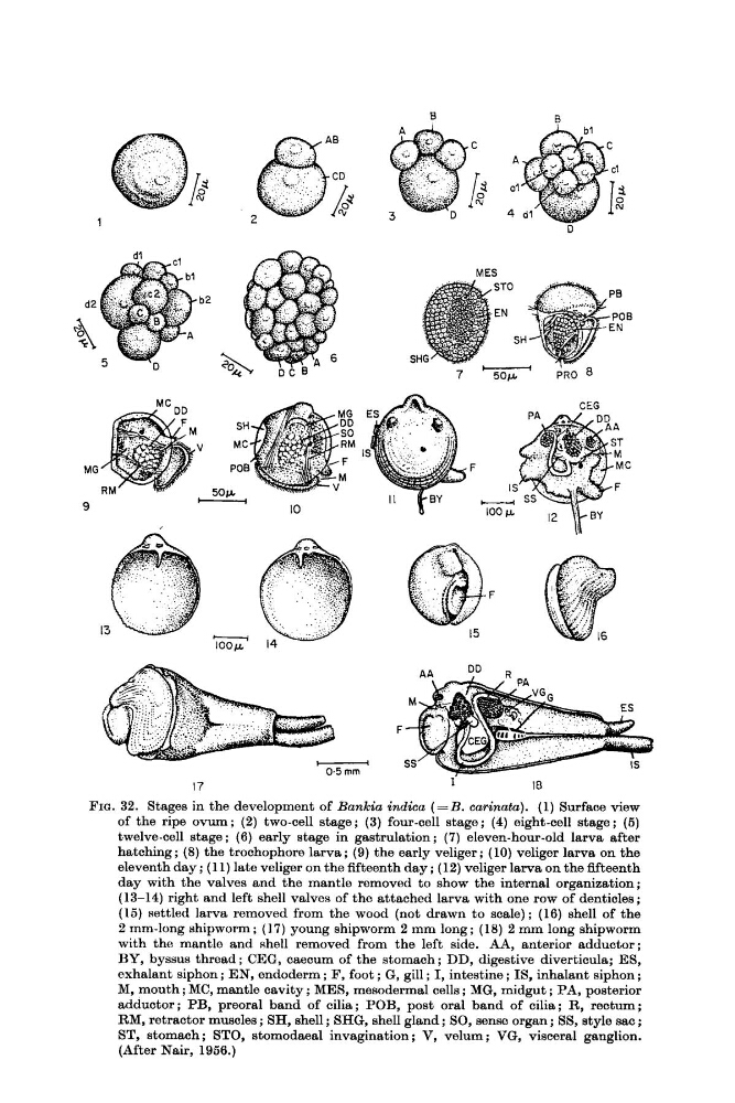
FIG. 32. Stages in the development of Bankia indica ( = B . carinata). (1) Surface view
of the ripe ovum; (2) two-cell stage; (3) four-cell stage; (4) eight-cell stage; (5)
twelve-cell stage ; (6) early stage in gastrulation; (7) eleven-hour-old larva after
hatching ; (8) the trochophore larva ; (9) the early veliger ; (10) veliger larva on the
eleventh day ; (1 1) late veliger on the fifteenth day ; (12) veliger larva on the fifteenth
day with the valves and the mantle removed to show the internal organization;
(13-14) right and left shell valves of the attached larva with one row of denticles ;
(15) settled larva removed from the wood (not drawn to scale) ; (16) shell of the
2 mm-long shipworm ; (17) young shipworm 2 mm long ; (18) 2 mm long shipworm
with the mantle and shell removed from the left side. AA, anterior adductor;
BY, byssus thread; CEG, caecum of the stomach; DD, digestive diverticula; ES,
exhalant siphon ; EN, endoderm ; F, foot ; G, gill ; I, intestine ; IS, inhalant siphon ;
M, mouth ; MC, mantle cavity ; MES, mesodermal cells ; MG, midgut ; PA, posterior
adductor; PB, preoral band of cilia; POB, post oral band of cilia; R, rectum;
RM, retractor muscles ; SH, shell ; SHG, shell gland ; SO, sense organ ; SS, style sac ;
ST, stomach ; STO, stomodaeal invagination ; V, velum ; VG, visceral ganglion.
(After Nair, 1956.)
R
1
C
A
O1
4
d2
B
%
C
MG
9
1 0
F
F
- ss
0 5 m m
AA
OD R
I'
17
FIG. 32. Stages in the development of Bankia indica ( = B . carinata). (1) Surface view
of the ripe ovum; (2) two-cell stage; (3) four-cell stage; (4) eight-cell stage; (5)
twelve-cell stage ; (6) early stage in gastrulation; (7) eleven-hour-old larva after
hatching ; (8) the trochophore larva ; (9) the early veliger ; (10) veliger larva on the
eleventh day ; (1 1) late veliger on the fifteenth day ; (12) veliger larva on the fifteenth
day with the valves and the mantle removed to show the internal organization;
(13-14) right and left shell valves of the attached larva with one row of denticles ;
(15) settled larva removed from the wood (not drawn to scale) ; (16) shell of the
2 mm-long shipworm ; (17) young shipworm 2 mm long ; (18) 2 mm long shipworm
with the mantle and shell removed from the left side. AA, anterior adductor;
BY, byssus thread; CEG, caecum of the stomach; DD, digestive diverticula; ES,
exhalant siphon ; EN, endoderm ; F, foot ; G, gill ; I, intestine ; IS, inhalant siphon ;
M, mouth ; MC, mantle cavity ; MES, mesodermal cells ; MG, midgut ; PA, posterior
adductor; PB, preoral band of cilia; POB, post oral band of cilia; R, rectum;
RM, retractor muscles ; SH, shell ; SHG, shell gland ; SO, sense organ ; SS, style sac ;
ST, stomach ; STO, stomodaeal invagination ; V, velum ; VG, visceral ganglion.
(After Nair, 1956.)