

388
N. BALAKRISHNAN NAIR AND M. SAFCASWATHY
12. The nervous system
The disposition of the various nerve centres and nerves in the larva,
the changes during growth and the nature of the system in adult Bankia
gouldi have been described by Sigerfoos (1908), who found that Teredo
navalis and T . dilatata conform to the same pattern. This is also true of
Bankia indica (Nair, 1955). The cerebral ganglia are distinct, rounded
bodies disposed dorso-lateral to the mouth behind the anterior adductor
muscle and connected by a short commissure. The pedal ganglion is a
single, compact body embedded within the tissues of the foot at its
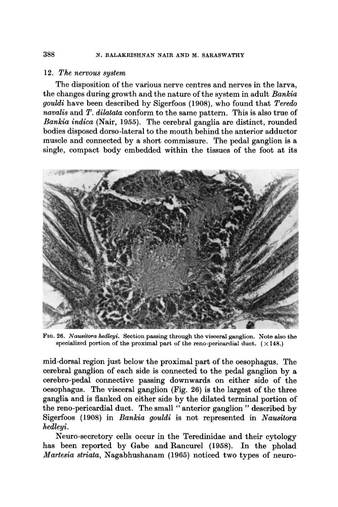
FIG. 26. Nausitora hedleyi. Section passing through the visceral ganglion. Note also the
specialized portion of the proximal part of the reno-pericardial duct. ( x 148.)
mid-dorsal region just below the proximal part of the oesophagus. The
cerebral ganglion of each side is connected to the pedal ganglion by a
cerebro-pedal connective passing downwards on either side of the
oesophagus. The visceral ganglion (Fig. 26) is the largest of the three
ganglia and is flanked on either side by the dilated terminal portion of
the reno-pericardial duct. The small " anterior ganglion ') described by
Sigerfoos (1908) in Bankia gouldi is not represented in Nausitora
hedleyi.
Neuro-secretory cells occur in the Teredinidae and their cytology
has been reported by Gabe and Rancurel (1958). In the pholad
Nartesia striata, Nagabhushanam (1965) noticed two types of neuro-
N. BALAKRISHNAN NAIR AND M. SAFCASWATHY
12. The nervous system
The disposition of the various nerve centres and nerves in the larva,
the changes during growth and the nature of the system in adult Bankia
gouldi have been described by Sigerfoos (1908), who found that Teredo
navalis and T . dilatata conform to the same pattern. This is also true of
Bankia indica (Nair, 1955). The cerebral ganglia are distinct, rounded
bodies disposed dorso-lateral to the mouth behind the anterior adductor
muscle and connected by a short commissure. The pedal ganglion is a
single, compact body embedded within the tissues of the foot at its
FIG. 26. Nausitora hedleyi. Section passing through the visceral ganglion. Note also the
specialized portion of the proximal part of the reno-pericardial duct. ( x 148.)
mid-dorsal region just below the proximal part of the oesophagus. The
cerebral ganglion of each side is connected to the pedal ganglion by a
cerebro-pedal connective passing downwards on either side of the
oesophagus. The visceral ganglion (Fig. 26) is the largest of the three
ganglia and is flanked on either side by the dilated terminal portion of
the reno-pericardial duct. The small " anterior ganglion ') described by
Sigerfoos (1908) in Bankia gouldi is not represented in Nausitora
hedleyi.
Neuro-secretory cells occur in the Teredinidae and their cytology
has been reported by Gabe and Rancurel (1958). In the pholad
Nartesia striata, Nagabhushanam (1965) noticed two types of neuro-