

382
N. BALAKRISHNAN NAIR AND M. SARASWATHY
the gill especially around the gill axis (Fig. 21). In Bankia gouldi,
Sigerfoos distinguished between spherical bodies and structures derived
from ramifying dentritic processes. Enlarged portions of the dentritic
processes may become surrounded by specialized epithelial cells derived
from those lining the gill or these cells may enclose a granular protoplasm with lightly staining nuclei and darkly staining rods projecting
towards blood spaces inside the gills. In Bankia indica, Nair (1957a)
recognized in the gill laminae two structures composed of cells differing
in histological details. A highly irregular ramification of filamentous
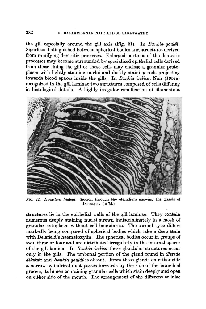
FIU. 22. Nausitora hedleyi. Section through the ctenidium showing the glands of
Deshayes. ( x 75.)
structures lie in the epithelial walls of the gill laminae. They contain
numerous deeply staining nuclei strewn indiscriminately in a mesh of
granular cytoplasm without cell boundaries. The second type differs
markedly being composed of spherical bodies which take a deep stain
with Delafield’s haematoxylin. The spherical bodies occur in groups of
two, three or four and are distributed irregularly in the internal spaces
of the gill lamina. In Bankia indica these glandular structures occur
only in the gills. The umbonal portion of the gland found in Teredo
dilatata and Bankia gouldi is absent. From these glands on either side
a narrow cylindrical duct passes forwards by the side of the branchial
groove, its lumen containing granular cells which stain deeply and open
on either side of the mouth. The arrangement of the different cellular
N. BALAKRISHNAN NAIR AND M. SARASWATHY
the gill especially around the gill axis (Fig. 21). In Bankia gouldi,
Sigerfoos distinguished between spherical bodies and structures derived
from ramifying dentritic processes. Enlarged portions of the dentritic
processes may become surrounded by specialized epithelial cells derived
from those lining the gill or these cells may enclose a granular protoplasm with lightly staining nuclei and darkly staining rods projecting
towards blood spaces inside the gills. In Bankia indica, Nair (1957a)
recognized in the gill laminae two structures composed of cells differing
in histological details. A highly irregular ramification of filamentous
FIU. 22. Nausitora hedleyi. Section through the ctenidium showing the glands of
Deshayes. ( x 75.)
structures lie in the epithelial walls of the gill laminae. They contain
numerous deeply staining nuclei strewn indiscriminately in a mesh of
granular cytoplasm without cell boundaries. The second type differs
markedly being composed of spherical bodies which take a deep stain
with Delafield’s haematoxylin. The spherical bodies occur in groups of
two, three or four and are distributed irregularly in the internal spaces
of the gill lamina. In Bankia indica these glandular structures occur
only in the gills. The umbonal portion of the gland found in Teredo
dilatata and Bankia gouldi is absent. From these glands on either side
a narrow cylindrical duct passes forwards by the side of the branchial
groove, its lumen containing granular cells which stain deeply and open
on either side of the mouth. The arrangement of the different cellular