

9. INTERNAL ANATOMY AND PHYSIOLOQY
261
heart and carry blood to the posterior regions of the ovary or to the vasa
deferentia. The two remaining vessels of the five originating from the
posterior end of the heart are the paired arteriae abdominalis superiores
(Fig. 96). These supply, by means of branches, the pleopods, abdominal
muscles and epithelium. The left arteria abdominalis superior ends in
the fourth abdominal segment (Fig. 96). I n the fifth abdominal segment,
the right arteria abdominalis superior supplies both members of the
fifth pair of pleopods as well as the tissues on both sides of the body.
The posterior part of this artery, by dichotomous branching, produces
a branch to each member of the two pairs of uropods and two branches
to the telson.
rnac
I \
/P
‘b
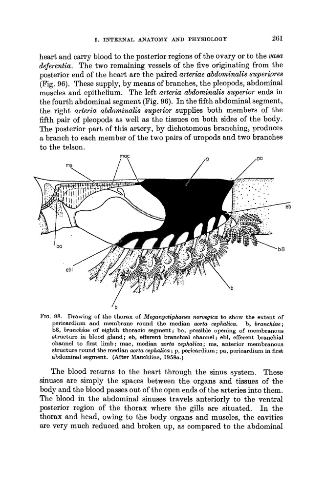
FIG. 98. Drawing of the thorax of Meganyctiphanes norvegica to show the extent of
pericardium and membrane round the median aorta cephalica. b, branchiae ;
b8, branchiae of eighth thoracic segment ; bo, possible opening of membranous
structure in blood gland ; eb, efferent branchial channel ; ebl, efferent branchial
channel to first limb; rnac, median aorta cephalica; ms, anterior membranous
structure round the median aorta cephalica ; p, pericardium ; pa, pericardium in first
abdominal segment. (After Mauchline, 1958a.)
The blood returns to the heart through the sinus system. These
sinuses are simply the spaces between the organs and tissues of the
body and the blood passes out of the open ends of the arteries into them.
The blood in the abdominal sinuses travels anteriorly to the ventral
posterior region of the thorax where the gills are situated. I n the
thorax and head, owing to the body organs and muscles, the cavities
are very much reduced and broken up, as compared to the abdominal
261
heart and carry blood to the posterior regions of the ovary or to the vasa
deferentia. The two remaining vessels of the five originating from the
posterior end of the heart are the paired arteriae abdominalis superiores
(Fig. 96). These supply, by means of branches, the pleopods, abdominal
muscles and epithelium. The left arteria abdominalis superior ends in
the fourth abdominal segment (Fig. 96). I n the fifth abdominal segment,
the right arteria abdominalis superior supplies both members of the
fifth pair of pleopods as well as the tissues on both sides of the body.
The posterior part of this artery, by dichotomous branching, produces
a branch to each member of the two pairs of uropods and two branches
to the telson.
rnac
I \
/P
‘b
FIG. 98. Drawing of the thorax of Meganyctiphanes norvegica to show the extent of
pericardium and membrane round the median aorta cephalica. b, branchiae ;
b8, branchiae of eighth thoracic segment ; bo, possible opening of membranous
structure in blood gland ; eb, efferent branchial channel ; ebl, efferent branchial
channel to first limb; rnac, median aorta cephalica; ms, anterior membranous
structure round the median aorta cephalica ; p, pericardium ; pa, pericardium in first
abdominal segment. (After Mauchline, 1958a.)
The blood returns to the heart through the sinus system. These
sinuses are simply the spaces between the organs and tissues of the
body and the blood passes out of the open ends of the arteries into them.
The blood in the abdominal sinuses travels anteriorly to the ventral
posterior region of the thorax where the gills are situated. I n the
thorax and head, owing to the body organs and muscles, the cavities
are very much reduced and broken up, as compared to the abdominal