

184
THE BIOLOGY OF EUPHAUSIIDS
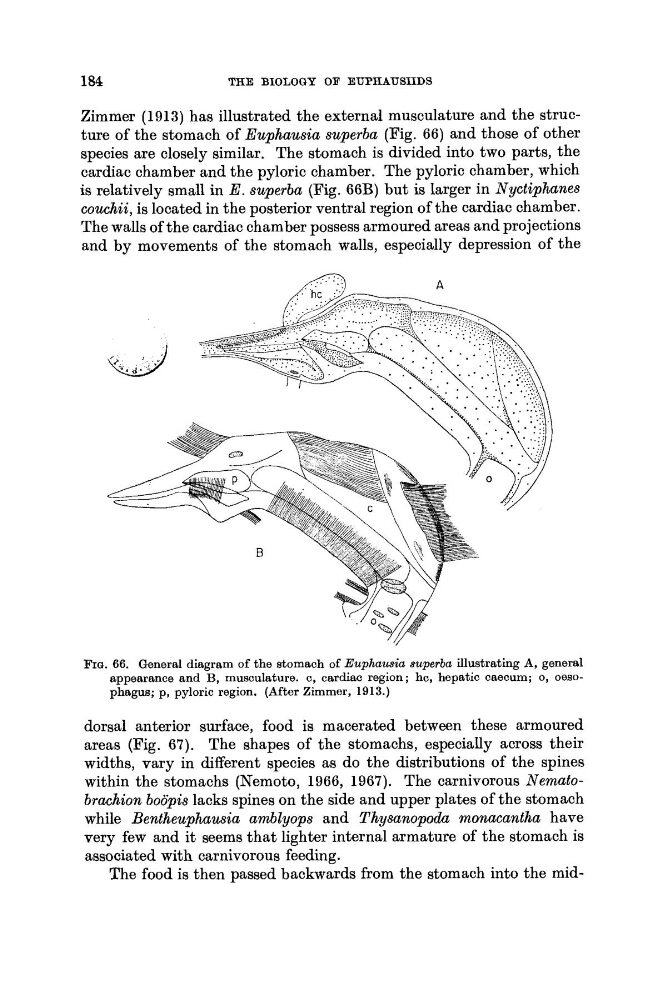
Zimmer (1913) has illustrated the external musculature and the structure of the stomach of Euphausia superba (Fig. 66) and those of other
species are closely similar. The stomach is divided into two parts, the
cardiac chamber and the pyloric chamber. The pyloric chamber, which
is relatively small in E . superba (Fig. 66B) but is larger in Nyctiphanes
couchii, is located in the posterior ventral region of the cardiac chamber.
The walls of the cardiac chamber possess armoured areas and projections
and by movements of the stomach walls, especially depression of the
FIG. 66. General diagram of the stomach of Euphausia superba illustrating A, general
appearance and B, musculature. c, cardiac region; hc, hepatic caecum; 0, oesophagus; p, pyloric region. (After Zimmer, 1913.)
dorsal anterior surface, food is macerated between these armoured
areas (Fig. 67). The shapes of the stomachs, especially across their
widths, vary in different species as do the distributions of the spines
within the stomachs (Nemoto, 1966, 1967). The carnivorous Nematobrachion boopis lacks spines on the side and upper plates of the stomach
while Bentheuphausia amblyops and Thysanopoda monacantha have
very few and it seems that lighter internal armature of the stomach is
associated with carnivorous feeding.
The food is then passed backwards from the stomach into the mid-
THE BIOLOGY OF EUPHAUSIIDS
Zimmer (1913) has illustrated the external musculature and the structure of the stomach of Euphausia superba (Fig. 66) and those of other
species are closely similar. The stomach is divided into two parts, the
cardiac chamber and the pyloric chamber. The pyloric chamber, which
is relatively small in E . superba (Fig. 66B) but is larger in Nyctiphanes
couchii, is located in the posterior ventral region of the cardiac chamber.
The walls of the cardiac chamber possess armoured areas and projections
and by movements of the stomach walls, especially depression of the
FIG. 66. General diagram of the stomach of Euphausia superba illustrating A, general
appearance and B, musculature. c, cardiac region; hc, hepatic caecum; 0, oesophagus; p, pyloric region. (After Zimmer, 1913.)
dorsal anterior surface, food is macerated between these armoured
areas (Fig. 67). The shapes of the stomachs, especially across their
widths, vary in different species as do the distributions of the spines
within the stomachs (Nemoto, 1966, 1967). The carnivorous Nematobrachion boopis lacks spines on the side and upper plates of the stomach
while Bentheuphausia amblyops and Thysanopoda monacantha have
very few and it seems that lighter internal armature of the stomach is
associated with carnivorous feeding.
The food is then passed backwards from the stomach into the mid-