

4. THE LARVAE
113
Zelikman, 196lb). Zimmer and Gruner (1956) suggest that the size of
the perivitelline space may be altered to change the specific gravity of
the eggs so that if the temperature and salinity characteristics of the
water mass in which they are laid changes, they can adapt to prevent
their sinking to the bottom. Similarly, this would cause variations in
embryo and capsule diameters between different geographical locations
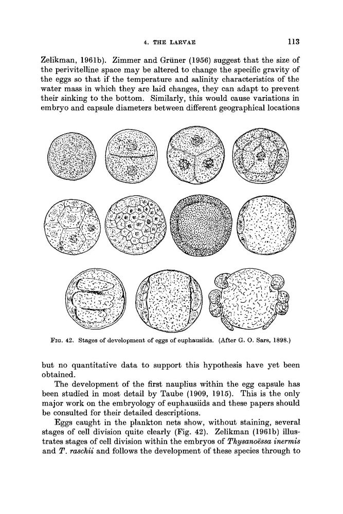
FIQ. 42. Stages of development of eggs of euphausiids. (After G . 0. Saw, 1898.)
but no quantitative data to support this hypothesis have yet been
obtained.
The development of the first nauplius within the egg capsule has
been studied in most detail by Taube (1909, 1915). This is the only
major work on the embryology of euphausiids and these papers should
be consulted for their detailed descriptions.
Eggs caught in the plankton nets show, without staining, several
stages of cell division quite clearly (Fig. 42). Zelikman (1961b) illustrates stages of cell division within the embryos of Thysanoessa inermis
and T . raschii and follows the development of these species through to
113
Zelikman, 196lb). Zimmer and Gruner (1956) suggest that the size of
the perivitelline space may be altered to change the specific gravity of
the eggs so that if the temperature and salinity characteristics of the
water mass in which they are laid changes, they can adapt to prevent
their sinking to the bottom. Similarly, this would cause variations in
embryo and capsule diameters between different geographical locations
FIQ. 42. Stages of development of eggs of euphausiids. (After G . 0. Saw, 1898.)
but no quantitative data to support this hypothesis have yet been
obtained.
The development of the first nauplius within the egg capsule has
been studied in most detail by Taube (1909, 1915). This is the only
major work on the embryology of euphausiids and these papers should
be consulted for their detailed descriptions.
Eggs caught in the plankton nets show, without staining, several
stages of cell division quite clearly (Fig. 42). Zelikman (1961b) illustrates stages of cell division within the embryos of Thysanoessa inermis
and T . raschii and follows the development of these species through to