

352
ELVEZIO GHIRARDELLI
damaged and the cut only affects the fin. Epithelial cells migrate
towards the wound, where many of them appear in mitosis ; they only
are responsible for the reconstitution of the fin rays and epidermal
glands (Ghirardelli, 1959a). The lateral fins also may be regenerated,
though more slowly, in 7-10 days. The caudal region sectioned at
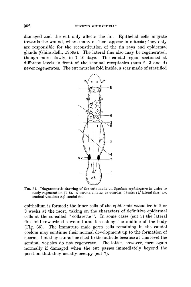
different levels in front of the seminal receptacles (cuts 2, 3 and 4)
never regenerates. The cut muscles fold inside, a scar made of stratified
FIG. 34. Diagrammatic drawing of the cuts made on Spadella cephnloptera in order to
study regeneration (1-8). cl corona ciliats ; ov ovaries ; t testes ; 2f lateral fins ; S. V.
seminal vesicles ; c.f. caudal fin.
epithelium is formed ; the inner cells of the epidermis vacuolize in 2 or
3 weeks a t the most, taking on the characters of definitive epidermal
cells at the so-called " collarette ',. I n some cases (cut 3) the lateral
fins fold towards the wound and fuse along the midline of the body
(Fig. 35). The immature male germ cells remaining in the caudal
coelom may continue their normal development up to the formation of
sperms, but they cannot be shed to the outside because at this level the
seminal vesicles do not regenerate. The latter, however, form again
normally if damaged when the cut passes immediately beyond the
position that they usually occupy (cut 7).
ELVEZIO GHIRARDELLI
damaged and the cut only affects the fin. Epithelial cells migrate
towards the wound, where many of them appear in mitosis ; they only
are responsible for the reconstitution of the fin rays and epidermal
glands (Ghirardelli, 1959a). The lateral fins also may be regenerated,
though more slowly, in 7-10 days. The caudal region sectioned at
different levels in front of the seminal receptacles (cuts 2, 3 and 4)
never regenerates. The cut muscles fold inside, a scar made of stratified
FIG. 34. Diagrammatic drawing of the cuts made on Spadella cephnloptera in order to
study regeneration (1-8). cl corona ciliats ; ov ovaries ; t testes ; 2f lateral fins ; S. V.
seminal vesicles ; c.f. caudal fin.
epithelium is formed ; the inner cells of the epidermis vacuolize in 2 or
3 weeks a t the most, taking on the characters of definitive epidermal
cells at the so-called " collarette ',. I n some cases (cut 3) the lateral
fins fold towards the wound and fuse along the midline of the body
(Fig. 35). The immature male germ cells remaining in the caudal
coelom may continue their normal development up to the formation of
sperms, but they cannot be shed to the outside because at this level the
seminal vesicles do not regenerate. The latter, however, form again
normally if damaged when the cut passes immediately beyond the
position that they usually occupy (cut 7).