

SOME ASPECTS O F T H E BIOLOGY OF THE CHAETOGNATHS
337
caused by the pressure of the eggs. When these enter the openings
(Fig. 28) they change their shape: they slip into the opening with one
pointed end which immediately after becomes larger, causing the
separation of the epithelial membrane from the underlying syncytium.
I n the cavity thus formed the egg continues to slip and thus the area of
separation becomes larger. The cavity so formed has rightly been
defined by Stevens as a temporary oviduct. As the eggs penetrate
between the epithelinm and the syncytium, they form a series of bags
a t different levels, each containing one or more eggs. When the eggs
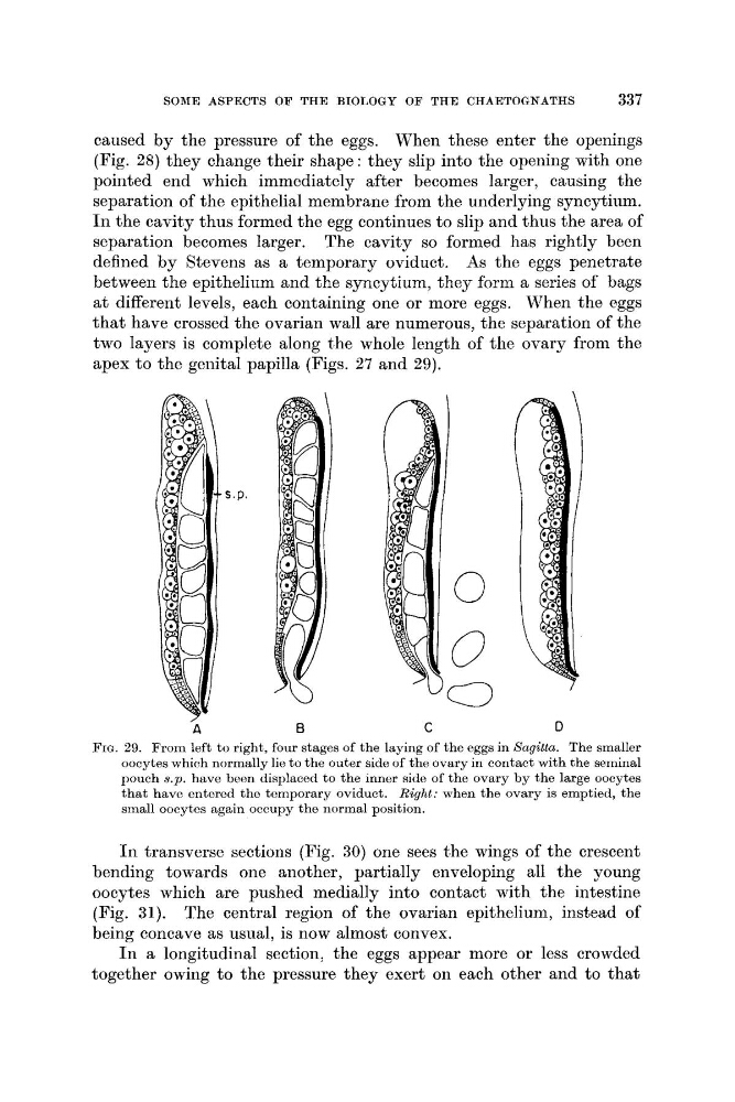
that have crossed the ovarian wall are numerous, the separation of the
two layers is complete along t'he whole length of the ovary from the
apex to the genital papilla (Figs. 27 and 29);
s.p.
/
A
D
FIG. 29. From left to right, four stages of the laying of the eggs in Sagitta. The smaller
oocytes which normally lie to the outer side of the ovary in contact with the seminal
pouch 8.p. have been displaced to the inner side of the ovary by the large oocytes
that have entered the temporary oviduct. Right: when the ovary is emptied, the
small oocytes again occupy the normal position.
I n transverse sections (Fig. 30) one sees t,he wings of the crescent
bending towards one another, partially enveloping all the young
oocytes which are pushed medially into contact with the intestine
(Fig. 31). The central region of the ovarian epithelium, instead of
being concave as usual, is now almost convex.
I n a longitudinal section, the eggs appear more or less crowded
together owing to the pressure they exert on each other and to that
337
caused by the pressure of the eggs. When these enter the openings
(Fig. 28) they change their shape: they slip into the opening with one
pointed end which immediately after becomes larger, causing the
separation of the epithelial membrane from the underlying syncytium.
I n the cavity thus formed the egg continues to slip and thus the area of
separation becomes larger. The cavity so formed has rightly been
defined by Stevens as a temporary oviduct. As the eggs penetrate
between the epithelinm and the syncytium, they form a series of bags
a t different levels, each containing one or more eggs. When the eggs
that have crossed the ovarian wall are numerous, the separation of the
two layers is complete along t'he whole length of the ovary from the
apex to the genital papilla (Figs. 27 and 29);
s.p.
/
A
D
FIG. 29. From left to right, four stages of the laying of the eggs in Sagitta. The smaller
oocytes which normally lie to the outer side of the ovary in contact with the seminal
pouch 8.p. have been displaced to the inner side of the ovary by the large oocytes
that have entered the temporary oviduct. Right: when the ovary is emptied, the
small oocytes again occupy the normal position.
I n transverse sections (Fig. 30) one sees t,he wings of the crescent
bending towards one another, partially enveloping all the young
oocytes which are pushed medially into contact with the intestine
(Fig. 31). The central region of the ovarian epithelium, instead of
being concave as usual, is now almost convex.
I n a longitudinal section, the eggs appear more or less crowded
together owing to the pressure they exert on each other and to that