

258
MARINE MOLLUSCS AS HOSTS FOR SYMBIOSES
representing developing myzorhynchus ; few elongate flask-shaped unicellular glands a t anterior end, in region of invagination ; parenchyma
filled with globular pale yellow bodies, each measuring 0.004-0.015 mm
x 0.004-0-01 1 mm ; few large granular cells (germinal cells) scattered
along periphery in posterior portion of body ; no typical cestode larval
hooks present. The detailed histology of this coracidium has been
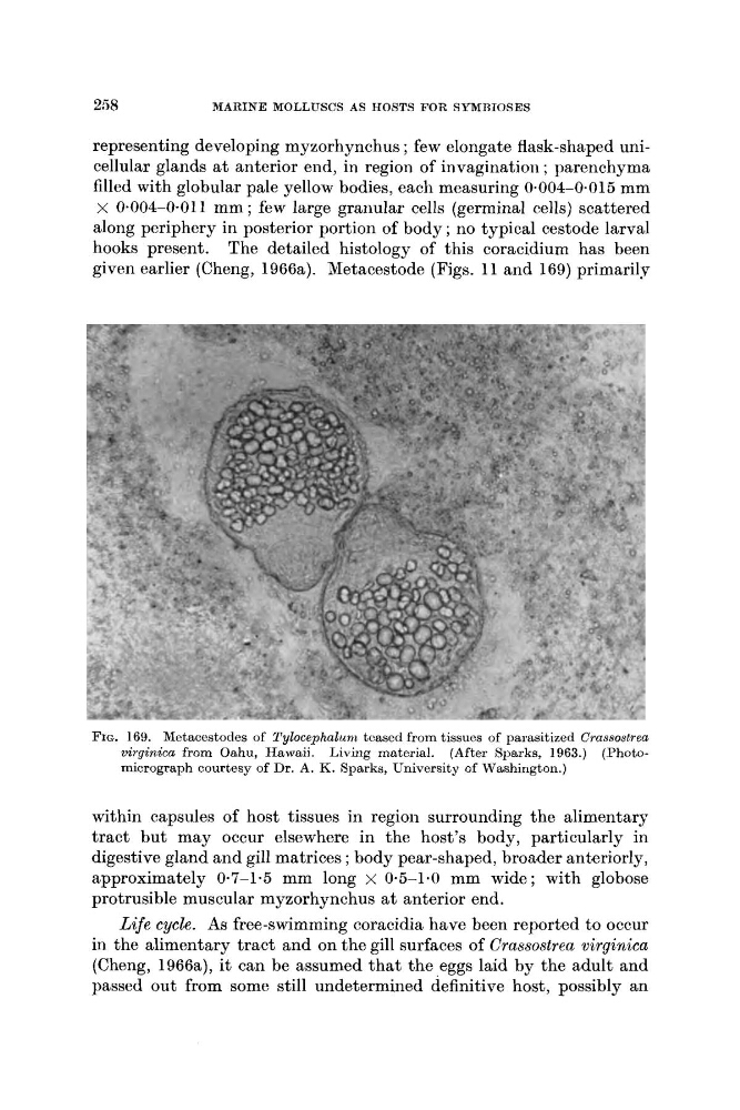
given earlier (Cheng, 1966a). Metacestode (Figs. 11 and 169) primarily
FIG. 169. Metacestodes of Tylocephalrm teased from tissues of parasitized Crassostrea
virginica from Oahu, Hawaii. Living material. (After Sparks, 1963.) (Photomicrograph courtesy of Dr. A. K. Sparks, University of Washington.)
within capsules of host tissues in region surrounding the alimentary
tract but may occur elsewhere in the host’s body, particularly in
digestive gland and gill matrices ; body pear-shaped, broader anteriorly,
approximately 0-7-1.5 mm long x 0-5-1.0 mm wide; with globose
protrusible muscular myzorhynchus a t anterior end.
Life cycle. As free-swimming coracidia have been reported to occur
in the alimentary tract and on the gill surfaces of Crassostrea virginica
(Cheng, 1966a), it can be assumed that the eggs laid by the adult and
passed out from some still undetermined definitive host, possibly an
MARINE MOLLUSCS AS HOSTS FOR SYMBIOSES
representing developing myzorhynchus ; few elongate flask-shaped unicellular glands a t anterior end, in region of invagination ; parenchyma
filled with globular pale yellow bodies, each measuring 0.004-0.015 mm
x 0.004-0-01 1 mm ; few large granular cells (germinal cells) scattered
along periphery in posterior portion of body ; no typical cestode larval
hooks present. The detailed histology of this coracidium has been
given earlier (Cheng, 1966a). Metacestode (Figs. 11 and 169) primarily
FIG. 169. Metacestodes of Tylocephalrm teased from tissues of parasitized Crassostrea
virginica from Oahu, Hawaii. Living material. (After Sparks, 1963.) (Photomicrograph courtesy of Dr. A. K. Sparks, University of Washington.)
within capsules of host tissues in region surrounding the alimentary
tract but may occur elsewhere in the host’s body, particularly in
digestive gland and gill matrices ; body pear-shaped, broader anteriorly,
approximately 0-7-1.5 mm long x 0-5-1.0 mm wide; with globose
protrusible muscular myzorhynchus a t anterior end.
Life cycle. As free-swimming coracidia have been reported to occur
in the alimentary tract and on the gill surfaces of Crassostrea virginica
(Cheng, 1966a), it can be assumed that the eggs laid by the adult and
passed out from some still undetermined definitive host, possibly an