

5. THE PHYLUM PROTOZOA
169
and Taylor (1966) have successfully demonstrated that the postplasmodial stages described by Couch et al. (1966) are antigenically
similar to the plasmodia1 stage generally recognized as that of M .
nelsoni and hence, until further evidences indicate otherwise, are
suspected to be stages in the life cycle of M . nelsoni. What Barrow
and Taylor did was to prepare a fluorecein-conjugated antibody from
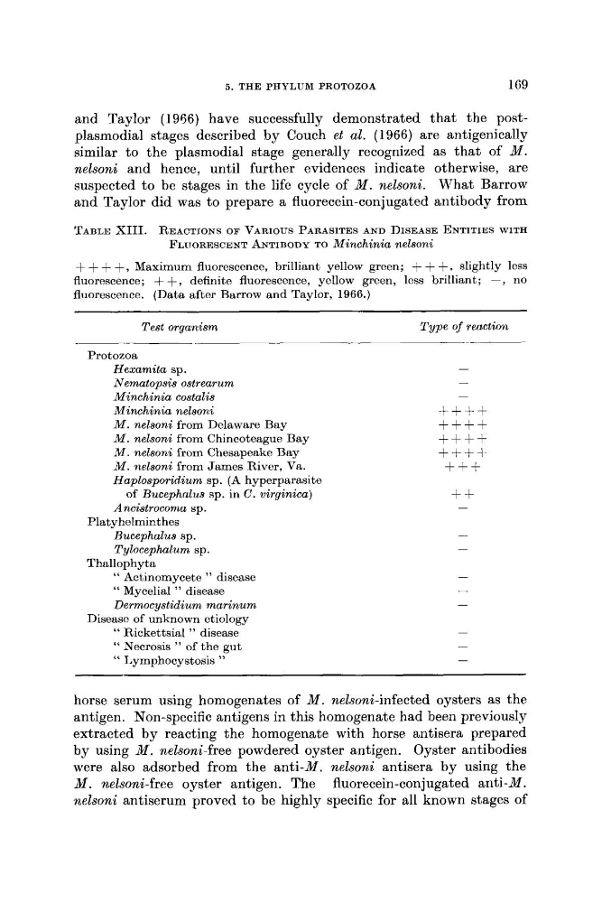
TABLE XIII. REACTIONS OF VARIOUS PARASITES AND DISEASE ENTITIES WITH
FLUORESCENT ANTIBODY TO Minchinia nelsoni
+ ~+ + +, Maximum fluorescence, brilliant yellow green; + + + , slightly less
fluorescence; + + , definite fluorescence, yellow green, less brilliant,; -, no
fluorescence. (Data after Barrow and Taylor, 1966.)
Test organism
Type of reaction
__
Protozoa
Hexamita sp.
Nematopsis ostrearum
Minchinia costalis
Minchink nelsoni
M . nelsoni from Delaware Bay
M . nelsoni from Chincoteague Bay
M . nelsoni from Chesapeake Bay
M . nelsoni from James River, Va.
Haplosporidium sp. (A hyperparasite
of Bucephrtlus sp. in C. virginica)
-
++++
++++
++++
++++
+++
+f
Ancistrocoma sp.
-
Bucephalus sp.
-
Tylocephalum sp.
-
" Actinomycetc " disease
-
" Mycelial " disease
-
Dermocystidium marinum
-
" Necrosis " of the gut
" Lymphocystosis "
-
Platyhelminthes
Thallophyta
Disease of unknown etiology
" Rickettsia1 " disease
-
-
horse serum using homogenates of M . nelsoni-infected oysters as the
antigen. Non-specific antigens in this homogenate had been previously
extracted by reacting the homogenate with horse antisera prepared
by using M . nelsoni-free powdered oyster antigen. Oyster antibodies
were also adsorbed from the anti-M. nelsoni antisera by using the
M. nelsoni-free oyster antigen. The fluorecein-conjugated anti-M.
nelsoni antiserum proved t o be highly specific for all known stages of
169
and Taylor (1966) have successfully demonstrated that the postplasmodial stages described by Couch et al. (1966) are antigenically
similar to the plasmodia1 stage generally recognized as that of M .
nelsoni and hence, until further evidences indicate otherwise, are
suspected to be stages in the life cycle of M . nelsoni. What Barrow
and Taylor did was to prepare a fluorecein-conjugated antibody from
TABLE XIII. REACTIONS OF VARIOUS PARASITES AND DISEASE ENTITIES WITH
FLUORESCENT ANTIBODY TO Minchinia nelsoni
+ ~+ + +, Maximum fluorescence, brilliant yellow green; + + + , slightly less
fluorescence; + + , definite fluorescence, yellow green, less brilliant,; -, no
fluorescence. (Data after Barrow and Taylor, 1966.)
Test organism
Type of reaction
__
Protozoa
Hexamita sp.
Nematopsis ostrearum
Minchinia costalis
Minchink nelsoni
M . nelsoni from Delaware Bay
M . nelsoni from Chincoteague Bay
M . nelsoni from Chesapeake Bay
M . nelsoni from James River, Va.
Haplosporidium sp. (A hyperparasite
of Bucephrtlus sp. in C. virginica)
-
++++
++++
++++
++++
+++
+f
Ancistrocoma sp.
-
Bucephalus sp.
-
Tylocephalum sp.
-
" Actinomycetc " disease
-
" Mycelial " disease
-
Dermocystidium marinum
-
" Necrosis " of the gut
" Lymphocystosis "
-
Platyhelminthes
Thallophyta
Disease of unknown etiology
" Rickettsia1 " disease
-
-
horse serum using homogenates of M . nelsoni-infected oysters as the
antigen. Non-specific antigens in this homogenate had been previously
extracted by reacting the homogenate with horse antisera prepared
by using M . nelsoni-free powdered oyster antigen. Oyster antibodies
were also adsorbed from the anti-M. nelsoni antisera by using the
M. nelsoni-free oyster antigen. The fluorecein-conjugated anti-M.
nelsoni antiserum proved t o be highly specific for all known stages of