

5 . THE PHYLUM PROTOZOA
157
stood how the individual merozoites comprising each gymnospore
become dispersed and subsequently become engulfed by other phagocytes. It is within phagocytes that the naked sporozoites are formed.
Other information. Although P. gigantea has been observed in the
process of attempting to penetrate the molluscan host’s gill epithelium,
no serious histopathological changes occur (Hatt, 1931). While within
phagocytes, no changes in the surrounding tissues are known to occur.
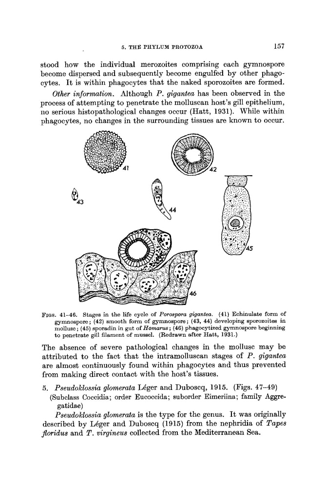
FIGS. 41-46. Stages in the life cycle of Porospora gigantea. (41) Echinulate form of
gymnospore; (42) smooth form of gymnospore; (43, 44) developing sporozoites in
mollusc ; (45) sporadin in gut of Hornarus; (46) phagocytized gymnospore beginning
to penetrate gill filament of mussel. (Redrawn after Hatt, 1931.)
The absence of severe pathological changes in the mollusc may be
attributed to the fact that the intramolluscan stages of P. gigantea
are almost continuously found within phagocytes and thus prevented
from making direct contact with the host’s tissues.
5. Pseudoklossia glomerata LBger and Duboscq, 1915. (Figs. 47-49)
(Subclass Coccidia; order Eucoccida; suborder Eimeriina; family Aggregat i d a e )
Pseudoklossia glomerata is the type for the genus. It was originally
described by LBger and Duboscq (1915) from the nephridia of Tapes
Jloridus and T . virgineus collected from the Mediterranean Sea.
157
stood how the individual merozoites comprising each gymnospore
become dispersed and subsequently become engulfed by other phagocytes. It is within phagocytes that the naked sporozoites are formed.
Other information. Although P. gigantea has been observed in the
process of attempting to penetrate the molluscan host’s gill epithelium,
no serious histopathological changes occur (Hatt, 1931). While within
phagocytes, no changes in the surrounding tissues are known to occur.
FIGS. 41-46. Stages in the life cycle of Porospora gigantea. (41) Echinulate form of
gymnospore; (42) smooth form of gymnospore; (43, 44) developing sporozoites in
mollusc ; (45) sporadin in gut of Hornarus; (46) phagocytized gymnospore beginning
to penetrate gill filament of mussel. (Redrawn after Hatt, 1931.)
The absence of severe pathological changes in the mollusc may be
attributed to the fact that the intramolluscan stages of P. gigantea
are almost continuously found within phagocytes and thus prevented
from making direct contact with the host’s tissues.
5. Pseudoklossia glomerata LBger and Duboscq, 1915. (Figs. 47-49)
(Subclass Coccidia; order Eucoccida; suborder Eimeriina; family Aggregat i d a e )
Pseudoklossia glomerata is the type for the genus. It was originally
described by LBger and Duboscq (1915) from the nephridia of Tapes
Jloridus and T . virgineus collected from the Mediterranean Sea.