

158
ERNEST SCHOFFENIELS
branes facing outside are permeable to Na and impervious to K, while
the contrary holds true for the membrane facing inside (104); (d) if a
microelectrode is introduced through the skin the potential difference
recorded between the solutions bathing the skin is established through
successive steps (one or two) (105); (e) lack of Κ in the inside solution
inhibits the active transport of Na, but a potassium-free outside medium
is without any effect (104).
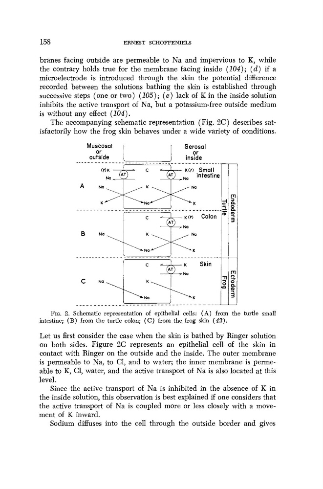
The accompanying schematic representation (Fig. 2C) describes satisfactorily how the frog skin behaves under a wide variety of conditions.
Muscosal
or
outside
Serosal
or
inside
Να ^
\
A
Na
Κ
J
^
^
κ
^^Na^^
^— κ(?) Small
V
N
a
intestine
Na
07
Endoderm
Β
Na
Κ
^^Na-*^
L— κ (?)
Colon
τ)
^
Na
07
Endoderm
C
Na
c
-J
Κ
^^Na
L— κ
Skin
ο
Ectoderm |
FIG. 2. Schematic representation of epithelial cells: (A) from the turtle small
intestine; (B) from the turtle colon; (C) from the frog skin (42).
Let us first consider the case when the skin is bathed by Ringer solution
on both sides. Figure 2C represents an epithelial cell of the skin in
contact with Ringer on the outside and the inside. The outer membrane
is permeable to Na, to CI, and to water; the inner membrane is permeable to K, CI, water, and the active transport of Na is also located at this
level.
Since the active transport of Na is inhibited in the absence of Κ in
the inside solution, this observation is best explained if one considers that
the active transport of Na is coupled more or less closely with a movement of Κ inward.
Sodium diffuses into the cell through the outside border and gives
ERNEST SCHOFFENIELS
branes facing outside are permeable to Na and impervious to K, while
the contrary holds true for the membrane facing inside (104); (d) if a
microelectrode is introduced through the skin the potential difference
recorded between the solutions bathing the skin is established through
successive steps (one or two) (105); (e) lack of Κ in the inside solution
inhibits the active transport of Na, but a potassium-free outside medium
is without any effect (104).
The accompanying schematic representation (Fig. 2C) describes satisfactorily how the frog skin behaves under a wide variety of conditions.
Muscosal
or
outside
Serosal
or
inside
Να ^
\
A
Na
Κ
J
^
^
κ
^^Na^^
^— κ(?) Small
V
N
a
intestine
Na
07
Endoderm
Β
Na
Κ
^^Na-*^
L— κ (?)
Colon
τ)
^
Na
07
Endoderm
C
Na
c
-J
Κ
^^Na
L— κ
Skin
ο
Ectoderm |
FIG. 2. Schematic representation of epithelial cells: (A) from the turtle small
intestine; (B) from the turtle colon; (C) from the frog skin (42).
Let us first consider the case when the skin is bathed by Ringer solution
on both sides. Figure 2C represents an epithelial cell of the skin in
contact with Ringer on the outside and the inside. The outer membrane
is permeable to Na, to CI, and to water; the inner membrane is permeable to K, CI, water, and the active transport of Na is also located at this
level.
Since the active transport of Na is inhibited in the absence of Κ in
the inside solution, this observation is best explained if one considers that
the active transport of Na is coupled more or less closely with a movement of Κ inward.
Sodium diffuses into the cell through the outside border and gives