

98
P. W. KENT
ton of cephalopods and in the radulae of Gastropods. It is also found in
the exoskeletons of most Ectoprocta and in Pogonophora.
Extensive reviews of the distribution of chitin over wide ranges of
animals are available, notably by the following authors: Biedermann
(39), Kunike (40), Campbell (41), Koch (42), Wiggles worth (14),
Richards (12) and Rudall (12a). Many surveys employed classical
methods of detection and, with the advent of more precise procedures,
confirmation seems to be very desirable in the majority of cases.
In the Arthropoda, chitin is not uniformly distributed throughout the
cuticle but is localized in the inner layer, the procuticle. The outer layer
(epicuticle) is invariably free from chitin. All arthropod cuticles exhibit
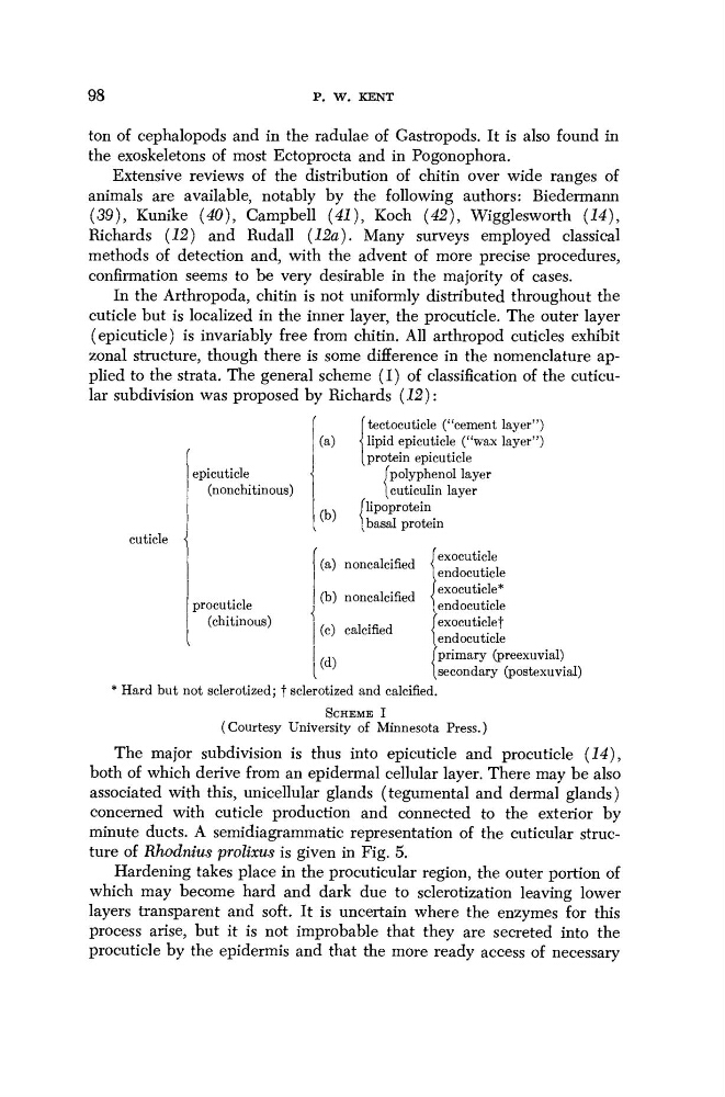
zonal structure, though there is some difference in the nomenclature applied to the strata. The general scheme (I) of classification of the cuticular subdivision was proposed by Richards (12):
cuticle
epicuticle
(nonchitinous)
(a)
(b)
[ tectocuticle ("cement layer")
Ί lipid epicuticle ("wax layer")
(protein epicuticle
(polyphenol layer
(cuticulin layer
(lipoprotein
[basal protein
(a) noncalcified
(b) noncalcified
(c) calcified
(d)
* Hard but not sclerotized; f sclerotized and calcified
procuticle
(chitinous)
exocuticle
endocuticle
exocuticle*
endocuticle
exocuticle f
endocuticle
primary (preexuvial)
secondary (postexuvial)
SCHEME I
(Courtesy University of Minnesota Press.)
The major subdivision is thus into epicuticle and procuticle (14),
both of which derive from an epidermal cellular layer. There may be also
associated with this, unicellular glands (tegumental and dermal glands)
concerned with cuticle production and connected to the exterior by
minute ducts. A semidiagrammatic representation of the cuticular structure of Rhodnius prolixus is given in Fig. 5.
Hardening takes place in the procuticular region, the outer portion of
which may become hard and dark due to sclerotization leaving lower
layers transparent and soft. It is uncertain where the enzymes for this
process arise, but it is not improbable that they are secreted into the
procuticle by the epidermis and that the more ready access of necessary
P. W. KENT
ton of cephalopods and in the radulae of Gastropods. It is also found in
the exoskeletons of most Ectoprocta and in Pogonophora.
Extensive reviews of the distribution of chitin over wide ranges of
animals are available, notably by the following authors: Biedermann
(39), Kunike (40), Campbell (41), Koch (42), Wiggles worth (14),
Richards (12) and Rudall (12a). Many surveys employed classical
methods of detection and, with the advent of more precise procedures,
confirmation seems to be very desirable in the majority of cases.
In the Arthropoda, chitin is not uniformly distributed throughout the
cuticle but is localized in the inner layer, the procuticle. The outer layer
(epicuticle) is invariably free from chitin. All arthropod cuticles exhibit
zonal structure, though there is some difference in the nomenclature applied to the strata. The general scheme (I) of classification of the cuticular subdivision was proposed by Richards (12):
cuticle
epicuticle
(nonchitinous)
(a)
(b)
[ tectocuticle ("cement layer")
Ί lipid epicuticle ("wax layer")
(protein epicuticle
(polyphenol layer
(cuticulin layer
(lipoprotein
[basal protein
(a) noncalcified
(b) noncalcified
(c) calcified
(d)
* Hard but not sclerotized; f sclerotized and calcified
procuticle
(chitinous)
exocuticle
endocuticle
exocuticle*
endocuticle
exocuticle f
endocuticle
primary (preexuvial)
secondary (postexuvial)
SCHEME I
(Courtesy University of Minnesota Press.)
The major subdivision is thus into epicuticle and procuticle (14),
both of which derive from an epidermal cellular layer. There may be also
associated with this, unicellular glands (tegumental and dermal glands)
concerned with cuticle production and connected to the exterior by
minute ducts. A semidiagrammatic representation of the cuticular structure of Rhodnius prolixus is given in Fig. 5.
Hardening takes place in the procuticular region, the outer portion of
which may become hard and dark due to sclerotization leaving lower
layers transparent and soft. It is uncertain where the enzymes for this
process arise, but it is not improbable that they are secreted into the
procuticle by the epidermis and that the more ready access of necessary