

2. CHITIN AND MUCOSUBSTANCES
95
sugar and to incorporate it into polysaccharides and other carbohydratecontaining polymers, there is little information concerning the presence
of chitin. Its presence in Saccharomycetes is still controversial. Greater
diversity exists among microorganisms in the number and variety of
amino sugars synthesized (16) in contrast to higher living forms. Not
only are D-glucosamine and D-galactosamine found, but also such sugars
as D-fucosamine (2-amino-2-deoxy-D-fucose), D-galactosaminouronic acid,
and neuraminic acids
(17-19).
These occur with other sugars generally as constituents of bacterial
polysaccharides (20, 21) which, though associated with the bacterial cell
wall, are not generally considered to be part of it. An amino sugar
polymer, colominic acid, synthesized by Escherichia coli K253, is comprised of N-acetylneuraminic acid residues glycosidically linked (22).
There is evidence of the presence of this material also in other species,
e.g., Citrobacter freundii. There is no morphological basis for comparison
of colominic acid with chitin, nor reason to associate it with the structure
of bacterial cell walls.
With the development of new methods of isolation of cell wall components (23), evidence suggests that gram-positive organisms, as in
Actinomycetales, possess a basal cell-wall membrane in which amino
sugars including muramic acid participate. This membrane may indeed
play a structural role analogous to that of chitin, but decisive evidence
on this is not yet available.
Elsewhere, there is increasing evidence (24) of the importance of
complex phosphorylated molecules, teichoic acids, in bacterial cell walls.
These substances, accounting for 40-60% of the cell wall of Lactobacillus
arabinosus, Bacillus subtilis, Staphylococcus aureus, and Staphylococcus
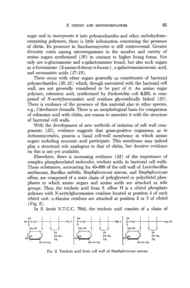
albus, are comprised of a main chain of polyglycerol or polyribitol phosphates to which amino sugars and amino acids are attached as side
groups. Thus, the teichoic acid from S. albus Η is a ribitol phosphate
polymer with N-acetylglucosamine residues located at position 4 of each
ribitol unit. D-Alanine residues are attached at position 2 or 3 of ribitol
(Fig. 2).
In S. lactis N.T.C.C. 7944, the teichoic acid consists of a chain of
OH
HO-P-O- HjCII
2
ο
CHj-OH
\OH
HO\
-CH a O
CO
CH- NI^
OH
•P-O- HjC-CH, Ο
CH- NHj
CH,
NH- CO- CH 3
OK
•POH a C-CHj-OH
FIG. 2. Teichoic acid from cell wall of Staphylococcus aureus.
95
sugar and to incorporate it into polysaccharides and other carbohydratecontaining polymers, there is little information concerning the presence
of chitin. Its presence in Saccharomycetes is still controversial. Greater
diversity exists among microorganisms in the number and variety of
amino sugars synthesized (16) in contrast to higher living forms. Not
only are D-glucosamine and D-galactosamine found, but also such sugars
as D-fucosamine (2-amino-2-deoxy-D-fucose), D-galactosaminouronic acid,
and neuraminic acids
(17-19).
These occur with other sugars generally as constituents of bacterial
polysaccharides (20, 21) which, though associated with the bacterial cell
wall, are not generally considered to be part of it. An amino sugar
polymer, colominic acid, synthesized by Escherichia coli K253, is comprised of N-acetylneuraminic acid residues glycosidically linked (22).
There is evidence of the presence of this material also in other species,
e.g., Citrobacter freundii. There is no morphological basis for comparison
of colominic acid with chitin, nor reason to associate it with the structure
of bacterial cell walls.
With the development of new methods of isolation of cell wall components (23), evidence suggests that gram-positive organisms, as in
Actinomycetales, possess a basal cell-wall membrane in which amino
sugars including muramic acid participate. This membrane may indeed
play a structural role analogous to that of chitin, but decisive evidence
on this is not yet available.
Elsewhere, there is increasing evidence (24) of the importance of
complex phosphorylated molecules, teichoic acids, in bacterial cell walls.
These substances, accounting for 40-60% of the cell wall of Lactobacillus
arabinosus, Bacillus subtilis, Staphylococcus aureus, and Staphylococcus
albus, are comprised of a main chain of polyglycerol or polyribitol phosphates to which amino sugars and amino acids are attached as side
groups. Thus, the teichoic acid from S. albus Η is a ribitol phosphate
polymer with N-acetylglucosamine residues located at position 4 of each
ribitol unit. D-Alanine residues are attached at position 2 or 3 of ribitol
(Fig. 2).
In S. lactis N.T.C.C. 7944, the teichoic acid consists of a chain of
OH
HO-P-O- HjCII
2
ο
CHj-OH
\OH
HO\
-CH a O
CO
CH- NI^
OH
•P-O- HjC-CH, Ο
CH- NHj
CH,
NH- CO- CH 3
OK
•POH a C-CHj-OH
FIG. 2. Teichoic acid from cell wall of Staphylococcus aureus.