

12. QUINONES: STRUCTURE AND DISTRIBUTION
669
O
O
O
O
Actinorhodin
Protoactinorhodin
(CXXV)
(CXXVI)
(2CH 3
RiRjjRgRt = < 2C02H
V.
C 8
H 16°2
from natural sources. When S. coelicolor is grown on a medium containing glycerol and glycine, the mycelium formed contains 15% (dry
weight) of pigment. This is normally isolated by alkaline extraction,
which gives a blue solution, but if the mycelium is harvested and
worked up under acid conditions, no blue color appears and a pale red
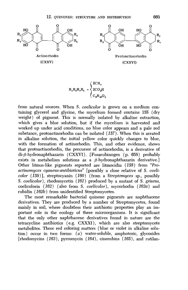
substance, protoactinorhodin can be isolated (157). When this is aerated
in alkaline solution, the initial yellow color quickly changes to blue,
with the formation of actinorhodin. This, and other evidence, shows
that protoactinorhodin, the precursor of actinorhodin, is a derivative of
di-ß-hydronaphthazarin (CXXVI). [Fusarubinogen (p. 658) probably
exists in metabolism solutions as a ß-hydronaphthazarin derivative.]
Other litmus-like pigments reported are litmocidin (158) from "Froactinomyces cyaneus-antibioticus" [possibly a close relative of S. coelicolor (159)], streptocyanin (160) (from a Streptomyces sp., possibly
S. coelicolor), rhodomycetin (161) produced by a mutant of S. griseus,
coelicolorin (162) (also from S. coelicolor), mycorhodin (162a) and
rubidin (162b) from unidentified Streptomycetes.
The most remarkable bacterial quinone pigments are naphthacene
derivatives. They are produced by a number of Streptomycetes, found
mainly in soil, where doubtless their antibiotic properties play an important role in the ecology of these microorganisms. It is significant
that the only other naphthacene derivatives found in nature are the
tetracycline antibiotics (e.g. CXXXI), which are also streptomycete
metabolites. These red coloring matters (blue or violet in alkaline solution) occur in two forms: (a) water-soluble, amphoteric, glycosides
[rhodomycins (163), pyrromycin (164), cinerubins (165), and rutilanHO
R 2 U^
HO