

296
KENNETH V. THIMANN AND GEORGE M. CURRY
due to complete compensation of the small light-growth reactions during
the 90 minute development period. The light-growth reactions observed
on symmetrical illumination by Went (53) and van Dillewijn (39)
substantiate this view; these workers observed definite light-growth reactions when the total energies and exposure times were much less than
those required to obtain base curvatures after 90 minutes. These lightgrowth reactions consisted of a growth rate deceleration followed by an
acceleration, the cycle being completed in 90 minutes. Similarly, Haig
(33) observed threshold base (curvature) responses at much lower
doses and after short exposures. Significantly, however, what he observed
was the first perceptible tilting of the coleoptile after the stimulus; this
occurs in less than 15 minutes, just as the decelerating light-growth reaction begins.
The above facts are consistent with the suggested mechanism. With
continuous unilateral exposures some A would always be in form B, and,
if there is a gradient of intensity across the organ, the level of A on the
exposed side will constantly be less than on the shaded side, accounting
for the large, continuously developing curvatures observed with such
exposures.
As far as the biochemistry of the conversion of A to B is concerned,
the only theoretical basis comes from the attempts to implicate riboflavin. Galston et al. (138) showed that catalase inhibits the destruction
of IAA by an oxidase preparation from pea stems, and that blue light
partly reverses this inhibition. The enzyme itself shows light activation
(139) which had been previously ascribed to the light reversal of the
effect of a natural inhibitor. They therefore proposed that IAA is
destroyed by a "light activable flavoprotein enzyme coupled through
H 2 0 2 to a peroxidase." However, Kenten (140) could find no trace of
flavoprotein in the preparation, and showed that the "IAA oxidase" consists of a peroxidase similar to that from horseradish (in agreement with
Galston et al.) plus a soluble heat-stable factor which is not a flavin
and which can be replaced by monophenols, aniline, resorcinol, or
maleic hydrazide, most of which are peroxidase substrates. The light
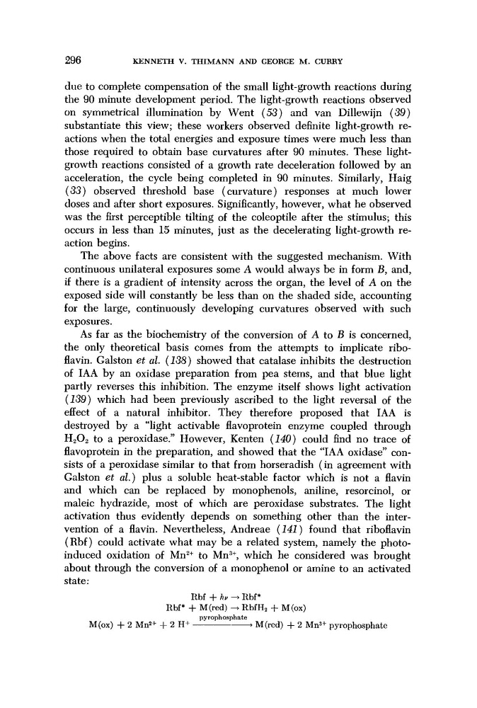
activation thus evidently depends on something other than the intervention of a flavin. Nevertheless, Andreae (141) found that riboflavin
(Rbf) could activate what may be a related system, namely the photoinduced oxidation of Mn
2+ to Mn
3+ , which he considered was brought
about through the conversion of a monophenol or amine to an activated
state:
Rbf + hv -> Rbf*
Rbf* + M(red) -> RbfH 2 + M(ox)
pyrophosphate
M(ox) + 2 Mn
2+ + 2 H+
> M(red) + 2 Mn
3+ pyrophosphate
KENNETH V. THIMANN AND GEORGE M. CURRY
due to complete compensation of the small light-growth reactions during
the 90 minute development period. The light-growth reactions observed
on symmetrical illumination by Went (53) and van Dillewijn (39)
substantiate this view; these workers observed definite light-growth reactions when the total energies and exposure times were much less than
those required to obtain base curvatures after 90 minutes. These lightgrowth reactions consisted of a growth rate deceleration followed by an
acceleration, the cycle being completed in 90 minutes. Similarly, Haig
(33) observed threshold base (curvature) responses at much lower
doses and after short exposures. Significantly, however, what he observed
was the first perceptible tilting of the coleoptile after the stimulus; this
occurs in less than 15 minutes, just as the decelerating light-growth reaction begins.
The above facts are consistent with the suggested mechanism. With
continuous unilateral exposures some A would always be in form B, and,
if there is a gradient of intensity across the organ, the level of A on the
exposed side will constantly be less than on the shaded side, accounting
for the large, continuously developing curvatures observed with such
exposures.
As far as the biochemistry of the conversion of A to B is concerned,
the only theoretical basis comes from the attempts to implicate riboflavin. Galston et al. (138) showed that catalase inhibits the destruction
of IAA by an oxidase preparation from pea stems, and that blue light
partly reverses this inhibition. The enzyme itself shows light activation
(139) which had been previously ascribed to the light reversal of the
effect of a natural inhibitor. They therefore proposed that IAA is
destroyed by a "light activable flavoprotein enzyme coupled through
H 2 0 2 to a peroxidase." However, Kenten (140) could find no trace of
flavoprotein in the preparation, and showed that the "IAA oxidase" consists of a peroxidase similar to that from horseradish (in agreement with
Galston et al.) plus a soluble heat-stable factor which is not a flavin
and which can be replaced by monophenols, aniline, resorcinol, or
maleic hydrazide, most of which are peroxidase substrates. The light
activation thus evidently depends on something other than the intervention of a flavin. Nevertheless, Andreae (141) found that riboflavin
(Rbf) could activate what may be a related system, namely the photoinduced oxidation of Mn
2+ to Mn
3+ , which he considered was brought
about through the conversion of a monophenol or amine to an activated
state:
Rbf + hv -> Rbf*
Rbf* + M(red) -> RbfH 2 + M(ox)
pyrophosphate
M(ox) + 2 Mn
2+ + 2 H+
> M(red) + 2 Mn
3+ pyrophosphate