

6. PHOTOTROPISM AND PHOTOTAXIS
279
that the sensitivity peak in the ultraviolet may reflect a particular structural modification of a carotenoid in the photoreceptor complex.
Bünning (108) found a close parallel between the distribution of
phototropic sensitivity and the distribution of carotenoids in the Avena
coleoptile, although most of the carotenoid lay in a region just below,
rather than at, the extreme tip, whereas Lange's data (38) imply that
the greatest sensitivity is in the topmost *4 mm. Because the work depended on histochemical observations, however, we have recently de0.800
0.600
0.400
0.200
0.000 350 400
450
500 550
600
650
700
Wavelength in m/i
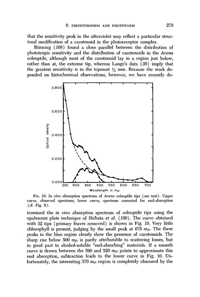
FIG. 10. In vivo absorption spectrum of Avena coleoptile tips (see text). Upper
curve, observed spectrum; lower curve, spectrum corrected for end-absorption
(cf. Fig. 8).
termined the in vivo absorption spectrum of coleoptile tips using the
opalescent plate technique of Shibata et al. (109). The curve obtained
with 32 tips (primary leaves removed) is shown in Fig. 10. Very little
chlorophyll is present, judging by the small peak at 675 πΐμ. The three
peaks in the blue region clearly show the presence of carotenoids. The
sharp rise below 500 τημ is partly attributable to scattering losses, but
in good part to alcohol-soluble "end-absorbing" materials. If a smooth
curve is drawn between the 390 and 520 τημ points to approximate this
end absorption, subtraction leads to the lower curve in Fig. 10. Unfortunately, the interesting 370 τημ region is completely obscured by the
279
that the sensitivity peak in the ultraviolet may reflect a particular structural modification of a carotenoid in the photoreceptor complex.
Bünning (108) found a close parallel between the distribution of
phototropic sensitivity and the distribution of carotenoids in the Avena
coleoptile, although most of the carotenoid lay in a region just below,
rather than at, the extreme tip, whereas Lange's data (38) imply that
the greatest sensitivity is in the topmost *4 mm. Because the work depended on histochemical observations, however, we have recently de0.800
0.600
0.400
0.200
0.000 350 400
450
500 550
600
650
700
Wavelength in m/i
FIG. 10. In vivo absorption spectrum of Avena coleoptile tips (see text). Upper
curve, observed spectrum; lower curve, spectrum corrected for end-absorption
(cf. Fig. 8).
termined the in vivo absorption spectrum of coleoptile tips using the
opalescent plate technique of Shibata et al. (109). The curve obtained
with 32 tips (primary leaves removed) is shown in Fig. 10. Very little
chlorophyll is present, judging by the small peak at 675 πΐμ. The three
peaks in the blue region clearly show the presence of carotenoids. The
sharp rise below 500 τημ is partly attributable to scattering losses, but
in good part to alcohol-soluble "end-absorbing" materials. If a smooth
curve is drawn between the 390 and 520 τημ points to approximate this
end absorption, subtraction leads to the lower curve in Fig. 10. Unfortunately, the interesting 370 τημ region is completely obscured by the