

DIFFERENTIATION OF KIDNEY MESENCHYME
263
from the establishment of inductor-mesenehyme contact, after which
overt differentiation can no longer be prevented by the drug (Jainchill
et al, 1964). The development of this resistance cannot be attributed to
reduced penetration of the drug, as seen from experiments employing
labeled actinomycin. Both determination of total incorporation and
radioautography studies indicate that the drug is incorporated into the
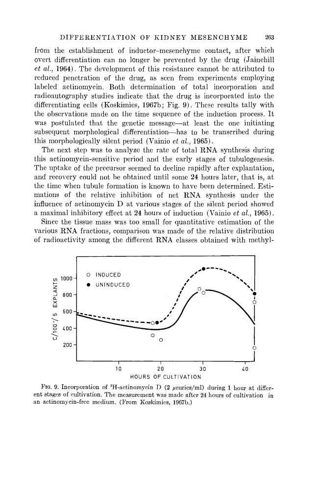
differentiating cells (Koskimies, 1967b; Fig. 9). These results tally with
the observations made on the time sequence of the induction process. It
was postulated that the genetic message—at least the one initiating
subsequent morphological differentiation—has to be transcribed during
this morphologically silent period (Vainio et al., 1965).
The next step was to analyze the rate of total RNA synthesis during
this actinomycin-sensitive period and the early stages of tubulogenesis.
The uptake of the precursor seemed to decline rapidly after explantation,
and recovery could not be obtained until some 24 hours later, that is, at
the time when tubule formation is known to have been determined. Estimations of the relative inhibition of net RNA synthesis under the
influence of actinomycin D at various stages of the silent period showed
a maximal inhibitory effect at 24 hours of induction (Vainio et al., 1965).
Since the tissue mass was too small for quantitative estimation of the
various RNA fractions, comparison was made of the relative distribution
of radioactivity among the different RNA classes obtained with methyln
1
1
r
10
20
30
40
HOURS OF CULTIVATION
FIG. 9. Incorporation of
3
H-actinomycin D (2 ^curies/ml) during 1 hour at different stages of cultivation. The measurement was made after 24 hours of cultivation in
an actinomycin-free medium. (From Koskimies, 1967b.)
C/l00"/5 EXPLANTS
263
from the establishment of inductor-mesenehyme contact, after which
overt differentiation can no longer be prevented by the drug (Jainchill
et al, 1964). The development of this resistance cannot be attributed to
reduced penetration of the drug, as seen from experiments employing
labeled actinomycin. Both determination of total incorporation and
radioautography studies indicate that the drug is incorporated into the
differentiating cells (Koskimies, 1967b; Fig. 9). These results tally with
the observations made on the time sequence of the induction process. It
was postulated that the genetic message—at least the one initiating
subsequent morphological differentiation—has to be transcribed during
this morphologically silent period (Vainio et al., 1965).
The next step was to analyze the rate of total RNA synthesis during
this actinomycin-sensitive period and the early stages of tubulogenesis.
The uptake of the precursor seemed to decline rapidly after explantation,
and recovery could not be obtained until some 24 hours later, that is, at
the time when tubule formation is known to have been determined. Estimations of the relative inhibition of net RNA synthesis under the
influence of actinomycin D at various stages of the silent period showed
a maximal inhibitory effect at 24 hours of induction (Vainio et al., 1965).
Since the tissue mass was too small for quantitative estimation of the
various RNA fractions, comparison was made of the relative distribution
of radioactivity among the different RNA classes obtained with methyln
1
1
r
10
20
30
40
HOURS OF CULTIVATION
FIG. 9. Incorporation of
3
H-actinomycin D (2 ^curies/ml) during 1 hour at different stages of cultivation. The measurement was made after 24 hours of cultivation in
an actinomycin-free medium. (From Koskimies, 1967b.)
C/l00"/5 EXPLANTS