

D I F F E R E N T I A T I O N O F K I D N E Y M E S E N C H Y M E
261
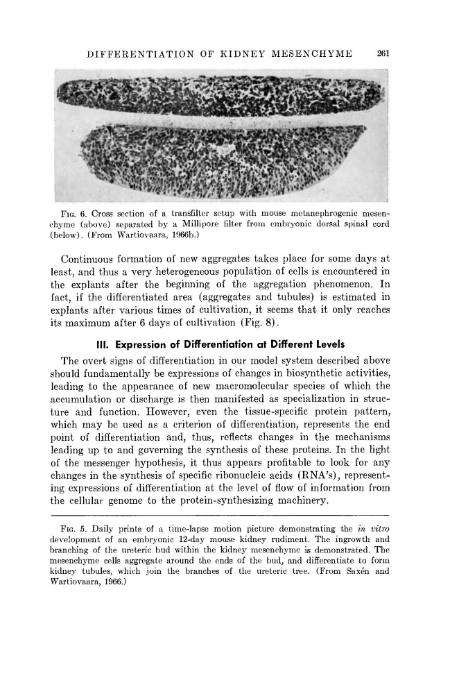
FIG. 6. Cross section of a transfilter setup with mouse metanephrogenic mesenchyme (above) separated by a Millipore filter from embryonic dorsal spinal cord
(below). (From Wartiovaara, 1966b.)
Continuous formation of new aggregates takes place for some days at
least, and thus a very heterogeneous population of cells is encountered in
the explants after the beginning of the aggregation phenomenon. In
fact, if the differentiated area (aggregates and tubules) is estimated in
explants after various times of cultivation, it seems that it only reaches
its maximum after 6 days of cultivation (Fig. 8 ) .
III. Expression of Differentiation at Different Levels
The overt signs of differentiation in our model system described above
should fundamentally be expressions of changes in biosynthetic activities,
leading to the appearance of new macromolecular species of which the
accumulation or discharge is then manifested as specialization in structure and function. However, even the tissue-specific protein pattern,
which m a y be used as a criterion of differentiation, represents the end
point of differentiation and, thus, reflects changes in the mechanisms
leading up to and governing the synthesis of these proteins. In the light
of the messenger hypothesis, it thus appears profitable to look for any
changes in the synthesis of specific ribonucleic acids ( R N A ' s ) , representing expressions of differentiation at the level of flow of information from
the cellular genome to the protein-synthesizing machinery.
FIG. 5. Daily prints of a time-lapse motion picture demonstrating the in vitro
development of an embryonic 12-day mouse kidney rudiment. The ingrowth and
branching of the ureteric bud within the kidney mesenchyme is demonstrated. The
mesenchyme cells aggregate around the ends of the bud, and differentiate to form
kidney tubules, which join the branches of the ureteric tree. (From Saxen and
Wartiovaara, 1966.)
261
FIG. 6. Cross section of a transfilter setup with mouse metanephrogenic mesenchyme (above) separated by a Millipore filter from embryonic dorsal spinal cord
(below). (From Wartiovaara, 1966b.)
Continuous formation of new aggregates takes place for some days at
least, and thus a very heterogeneous population of cells is encountered in
the explants after the beginning of the aggregation phenomenon. In
fact, if the differentiated area (aggregates and tubules) is estimated in
explants after various times of cultivation, it seems that it only reaches
its maximum after 6 days of cultivation (Fig. 8 ) .
III. Expression of Differentiation at Different Levels
The overt signs of differentiation in our model system described above
should fundamentally be expressions of changes in biosynthetic activities,
leading to the appearance of new macromolecular species of which the
accumulation or discharge is then manifested as specialization in structure and function. However, even the tissue-specific protein pattern,
which m a y be used as a criterion of differentiation, represents the end
point of differentiation and, thus, reflects changes in the mechanisms
leading up to and governing the synthesis of these proteins. In the light
of the messenger hypothesis, it thus appears profitable to look for any
changes in the synthesis of specific ribonucleic acids ( R N A ' s ) , representing expressions of differentiation at the level of flow of information from
the cellular genome to the protein-synthesizing machinery.
FIG. 5. Daily prints of a time-lapse motion picture demonstrating the in vitro
development of an embryonic 12-day mouse kidney rudiment. The ingrowth and
branching of the ureteric bud within the kidney mesenchyme is demonstrated. The
mesenchyme cells aggregate around the ends of the bud, and differentiate to form
kidney tubules, which join the branches of the ureteric tree. (From Saxen and
Wartiovaara, 1966.)