

188
PAUL BRIEN
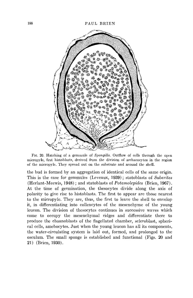
FIG. 20. Hatching of a gemmule of Spongilla. Outflow of cells through the open
micropyle, first histoblasts, derived from the division of archaeocytes in the region
of the micropyle. They spread out on the substrate and around the shell.
the bud is formed by an aggregation of identical cells of the same origin.
This is the case for gemmules (Leveaux, 1939); statoblasts of Suberites
(Herlant-Meewis, 1948); and statoblasts of Potamolepides
(Brien, 1967).
At the time of germination, the thesocytes divide along the axis of
polarity to give rise to histoblasts. The first to appear are those nearest
to the micropyle. They are, thus, the first to leave the shell to envelop
it, in differentiating into collencytes of the mesenchyme of the young
leucon. The division of thesocytes continues in successive waves which
come to occupy the mesenchymal ridges and differentiate there to
produce the choanoblasts of the flagellated chamber, scleroblast, spherical cells, amebocytes. Just when the young leucon has all its components,
the water-circulating system is laid out, formed, and prolonged to the
osculum. The small sponge is established and functional (Figs. 20 and
21) (Brien, 1930).
PAUL BRIEN
FIG. 20. Hatching of a gemmule of Spongilla. Outflow of cells through the open
micropyle, first histoblasts, derived from the division of archaeocytes in the region
of the micropyle. They spread out on the substrate and around the shell.
the bud is formed by an aggregation of identical cells of the same origin.
This is the case for gemmules (Leveaux, 1939); statoblasts of Suberites
(Herlant-Meewis, 1948); and statoblasts of Potamolepides
(Brien, 1967).
At the time of germination, the thesocytes divide along the axis of
polarity to give rise to histoblasts. The first to appear are those nearest
to the micropyle. They are, thus, the first to leave the shell to envelop
it, in differentiating into collencytes of the mesenchyme of the young
leucon. The division of thesocytes continues in successive waves which
come to occupy the mesenchymal ridges and differentiate there to
produce the choanoblasts of the flagellated chamber, scleroblast, spherical cells, amebocytes. Just when the young leucon has all its components,
the water-circulating system is laid out, formed, and prolonged to the
osculum. The small sponge is established and functional (Figs. 20 and
21) (Brien, 1930).