

D E V E L O P M E N T S I N SEXUAL O R G A N O G E N E S I S
309
A second method of approaching the problem of the biosynthesis of
steroids is by the use of histochemical techniques to demonstrate the
presence of enzyme systems. These have been detected in the gonads,
interrenal tissue, or both.
(i) Elasmobranch fishes (Scyliorhinus stellaris). Well before hatching the
interrenal gland contains A
5
-3ß-hydroxysteroid dehydrogenase (Chieffi,
1965).
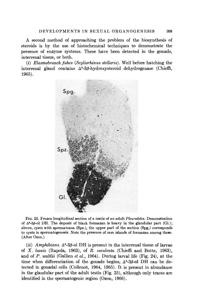
FIG. 25. Frozen longitudinal section of a testis of an adult Pleurodehs. Demonstration
of Δ
5
-3β-ο1 DH. The deposit of black formazan is heavy in the glandular part (Gl.);
above, cysts with spermatozoa (Spz.), the upper part of the section (Spg.) corresponds
to cysts in spermatogenesis. Note the presence of rare islands of formazan among these.
(After Ozon.)
(ii) Amphibians. Δ
5
-3β-ο1 DH is present in the interrenal tissue of larvae
of X. laevis (Rapola, 1963), of R. esculenta (Chieffi and Botte, 1963),
and of P. waltlii (Gallien et al., 1964). During larval life (Fig. 24), at the
time when differentiation of the gonads begins, Δ
5
-30-ο1 DH can be detected in gonadal cells (Collenot, 1964, 1965). It is present in abundance
in the glandular part of the adult testis (Fig. 25), although only traces are
identified in the spermatogenic region (Ozon, 1966).
309
A second method of approaching the problem of the biosynthesis of
steroids is by the use of histochemical techniques to demonstrate the
presence of enzyme systems. These have been detected in the gonads,
interrenal tissue, or both.
(i) Elasmobranch fishes (Scyliorhinus stellaris). Well before hatching the
interrenal gland contains A
5
-3ß-hydroxysteroid dehydrogenase (Chieffi,
1965).
FIG. 25. Frozen longitudinal section of a testis of an adult Pleurodehs. Demonstration
of Δ
5
-3β-ο1 DH. The deposit of black formazan is heavy in the glandular part (Gl.);
above, cysts with spermatozoa (Spz.), the upper part of the section (Spg.) corresponds
to cysts in spermatogenesis. Note the presence of rare islands of formazan among these.
(After Ozon.)
(ii) Amphibians. Δ
5
-3β-ο1 DH is present in the interrenal tissue of larvae
of X. laevis (Rapola, 1963), of R. esculenta (Chieffi and Botte, 1963),
and of P. waltlii (Gallien et al., 1964). During larval life (Fig. 24), at the
time when differentiation of the gonads begins, Δ
5
-30-ο1 DH can be detected in gonadal cells (Collenot, 1964, 1965). It is present in abundance
in the glandular part of the adult testis (Fig. 25), although only traces are
identified in the spermatogenic region (Ozon, 1966).