

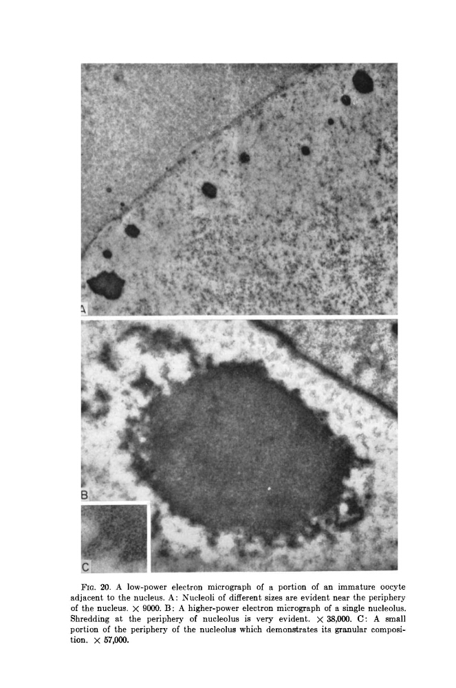
FIG. 20. A low-power electron micrograph of a portion of an immature oocyte
adjacent to the nucleus. A: Nucleoli of different sizes are evident near the periphery
of the nucleus. X 9000. B : A higher-power electron micrograph of a single nucleolus.
Shredding at the periphery of nucleolus is very evident, χ 38,000. C: A small
portion of the periphery of the nucleolus which demonstrates its granular composition. X 57,000.
adjacent to the nucleus. A: Nucleoli of different sizes are evident near the periphery
of the nucleus. X 9000. B : A higher-power electron micrograph of a single nucleolus.
Shredding at the periphery of nucleolus is very evident, χ 38,000. C: A small
portion of the periphery of the nucleolus which demonstrates its granular composition. X 57,000.