

U L T R A S T R U C T U R E OF T H E A M P H I B I A N E G G
187
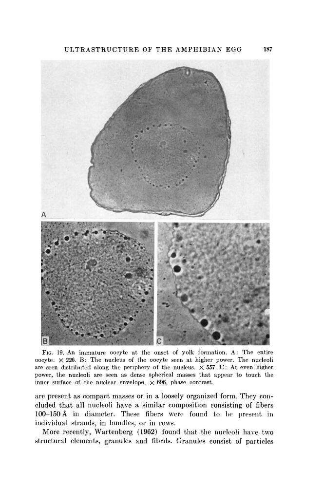
FIG. 19. An immature oocyte at the onset of yolk formation. A: The entire
oocyte. X 226. B : The nucleus of the oocyte seen at higher power. The nucleoli
are seen distributed along the periphery of the nucleus. X 557. C : At even higher
power, the nucleoli are seen as dense spherical masses that appear to touch the
inner surface of the nuclear envelope. X 696, phase contrast.
are present as compact masses or in a loosely organized form. They concluded that all nucleoli have a similar composition consisting of fibers
100-150 Ä in diameter. These fibers were found to be present in
individual strands, in bundles, or in rows.
More recently, Wartenberg (1962) found that the nucleoli have two
structural elements, granules and fibrils. Granules consist of particles
187
FIG. 19. An immature oocyte at the onset of yolk formation. A: The entire
oocyte. X 226. B : The nucleus of the oocyte seen at higher power. The nucleoli
are seen distributed along the periphery of the nucleus. X 557. C : At even higher
power, the nucleoli are seen as dense spherical masses that appear to touch the
inner surface of the nuclear envelope. X 696, phase contrast.
are present as compact masses or in a loosely organized form. They concluded that all nucleoli have a similar composition consisting of fibers
100-150 Ä in diameter. These fibers were found to be present in
individual strands, in bundles, or in rows.
More recently, Wartenberg (1962) found that the nucleoli have two
structural elements, granules and fibrils. Granules consist of particles