

178
SAUL W I S C H N I T Z E R
Evidence regarding the macromolecular structure of the nuclear envelope has been deduced from polarization microscopy. Schmidt (1939)
noticed birefringence in the "nuclear membrane" in freshly isolated
amphibian oocytes. Subsequently, Callan (1948) studied the isolated
nucleus with an improved optical system, and he reported that the
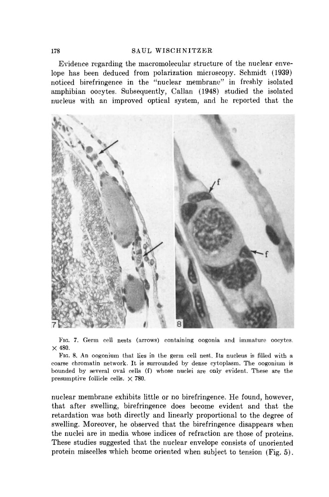
FIG. 7. Germ cell nests (arrows) containing oogonia and immature oocytes.
X 480.
FIG. 8. An oogonium that lies in the germ cell nest. Its nucleus is filled with a
coarse chromatin network. I t is surrounded by dense cytoplasm. The oogonium is
bounded by several oval cells (f) whose nuclei are only evident. These are the
presumptive follicle cells. X 780.
nuclear membrane exhibits little or no birefringence. He found, however,
that after swelling, birefringence does become evident and that the
retardation was both directly and linearly proportional to the degree of
swelling. Moreover, he observed that the birefringence disappears when
the nuclei are in media whose indices of refraction are those of proteins.
These studies suggested that the nuclear envelope consists of unoriented
protein miscelles which bcome oriented when subject to tension (Fig. 5).
SAUL W I S C H N I T Z E R
Evidence regarding the macromolecular structure of the nuclear envelope has been deduced from polarization microscopy. Schmidt (1939)
noticed birefringence in the "nuclear membrane" in freshly isolated
amphibian oocytes. Subsequently, Callan (1948) studied the isolated
nucleus with an improved optical system, and he reported that the
FIG. 7. Germ cell nests (arrows) containing oogonia and immature oocytes.
X 480.
FIG. 8. An oogonium that lies in the germ cell nest. Its nucleus is filled with a
coarse chromatin network. I t is surrounded by dense cytoplasm. The oogonium is
bounded by several oval cells (f) whose nuclei are only evident. These are the
presumptive follicle cells. X 780.
nuclear membrane exhibits little or no birefringence. He found, however,
that after swelling, birefringence does become evident and that the
retardation was both directly and linearly proportional to the degree of
swelling. Moreover, he observed that the birefringence disappears when
the nuclei are in media whose indices of refraction are those of proteins.
These studies suggested that the nuclear envelope consists of unoriented
protein miscelles which bcome oriented when subject to tension (Fig. 5).