

86
G.
REVERBERI
a yellow-orange fluorescence (De Vincentiis, 1959). The quantitative
spectrophotometric analysis of their fluorescence is represented by a
curve with two maxima, at 5800 Á and 5250 Á, which would indicate
the presence of a lipidie substance. In any case, the removal of the egg
envelopes does not interfere with a normal development.
2. The Cortex and Experiments on Localized Extirpation
The egg is limited by a cortex which, at fertilization, changes physically
and chemically. The physical changes can be demonstrated by a simple
50
51
52
53
54
55
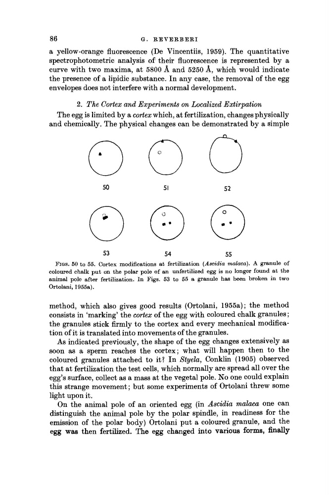
FIGS. 50 to 55. Cortex modifications at fertilization (Ascidia malaca). A granule of
coloured chalk put on the polar pole of an unfertilized egg is no longer found at the
animal pole after fertilization. In Figs. 53 to 55 a granule has been broken in two
Ortolani, 1955a).
method, which also gives good results (Ortolani, 1955a); the method
consists in 'marking' the cortex of the egg with coloured chalk granules;
the granules stick firmly to the cortex and every mechanical modification of it is translated into movements of the granules.
As indicated previously, the shape of the egg changes extensively as
soon as a sperm reaches the cortex; what will happen then to the
coloured granules attached to it? In Styela, Conklin (1905) observed
that at fertilization the test cells, which normally are spread all over the
egg's surface, collect as a mass at the vegetal pole. No one could explain
this strange movement; but some experiments of Ortolani threw some
light upon it.
On the animal pole of an oriented egg (in Ascidia malaca one can
distinguish the animal pole by the polar spindle, in readiness for the
emission of the polar body) Ortolani put a coloured granule, and the
egg was then fertilized. The egg changed into various forms, finally
G.
REVERBERI
a yellow-orange fluorescence (De Vincentiis, 1959). The quantitative
spectrophotometric analysis of their fluorescence is represented by a
curve with two maxima, at 5800 Á and 5250 Á, which would indicate
the presence of a lipidie substance. In any case, the removal of the egg
envelopes does not interfere with a normal development.
2. The Cortex and Experiments on Localized Extirpation
The egg is limited by a cortex which, at fertilization, changes physically
and chemically. The physical changes can be demonstrated by a simple
50
51
52
53
54
55
FIGS. 50 to 55. Cortex modifications at fertilization (Ascidia malaca). A granule of
coloured chalk put on the polar pole of an unfertilized egg is no longer found at the
animal pole after fertilization. In Figs. 53 to 55 a granule has been broken in two
Ortolani, 1955a).
method, which also gives good results (Ortolani, 1955a); the method
consists in 'marking' the cortex of the egg with coloured chalk granules;
the granules stick firmly to the cortex and every mechanical modification of it is translated into movements of the granules.
As indicated previously, the shape of the egg changes extensively as
soon as a sperm reaches the cortex; what will happen then to the
coloured granules attached to it? In Styela, Conklin (1905) observed
that at fertilization the test cells, which normally are spread all over the
egg's surface, collect as a mass at the vegetal pole. No one could explain
this strange movement; but some experiments of Ortolani threw some
light upon it.
On the animal pole of an oriented egg (in Ascidia malaca one can
distinguish the animal pole by the polar spindle, in readiness for the
emission of the polar body) Ortolani put a coloured granule, and the
egg was then fertilized. The egg changed into various forms, finally