

IX. DEVELOPMENT OF THE TELEOSTEAN EGG
419
Teleosts. The cells of the enveloping layer detach themselves separately
at the level of the lips; the continuity of the germinal surface is thus
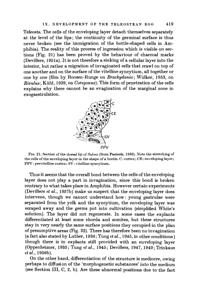
never broken (see the immigration of the bottle-shaped cells in Amphibia). The reality of this process of ingression which is visible on sections (Fig. 21) has been proved by the behaviour of charcoal marks
(Devillers, 1951a). It is not therefore a sinking of a cellular layer into the
interior, but rather a migration of invaginated cells that crawl on top of
one another and on the surface of the vitelline syncytium, all together or
one by one (film by Roosen-Runge on Brachydanio; Wülker, 1953, on
Rivulus; Kühl, 1939, on Goregonus). This form of penetration of the cells
explains why there cannot be an evagination of the marginal zone in
exogaestrulation.
FIG. 21. Section of the dorsal lip of Salmo (from Pasteéis, 1936). Note the stretching of
the cells of the enveloping layer in the shape of a bottle. C: cortex; CE: enveloping layer;
PPV: pervitelline cortex; SV: vitelline syncytium.
Thus it seems that the overall bond between the cells of the enveloping
layer does not play a part in invagination, since this bond is broken
contrary to what takes place in Amphibia. However certain experiments
(Devillers et al., 1957b) make us suspect that the enveloping layer does
intervene, though we cannot understand how: young gastrulae were
separated from the yolk and the syncytium, the enveloping layer was
scraped away and the germs put into cultivation (simplified White's
solution). The layer did not regenerate. In some cases the explants
differentiated at least some chorda and somites, but these structures
stay in very nearly the same surface positions they occupied in the plan
of presumptive areas (Fig. 22). There has therefore been no invagination
(a fact also stated by Luther, 1936; Tung et al., 1945, in other conditions)
though there is in explants still provided with an enveloping layer
(Oppenheimer, 1935; Tung et al, 1945; Devillers, 1947, 1949; Trinkaus
et al, 1956b).
On the other hand, differentiation of the structure is mediocre, owing
perhaps to diffusion of the 'morphogenetic substances' into the medium
(see Section III, C, 2, b). Are these abnormal positions due to the fact
CE
PFV
419
Teleosts. The cells of the enveloping layer detach themselves separately
at the level of the lips; the continuity of the germinal surface is thus
never broken (see the immigration of the bottle-shaped cells in Amphibia). The reality of this process of ingression which is visible on sections (Fig. 21) has been proved by the behaviour of charcoal marks
(Devillers, 1951a). It is not therefore a sinking of a cellular layer into the
interior, but rather a migration of invaginated cells that crawl on top of
one another and on the surface of the vitelline syncytium, all together or
one by one (film by Roosen-Runge on Brachydanio; Wülker, 1953, on
Rivulus; Kühl, 1939, on Goregonus). This form of penetration of the cells
explains why there cannot be an evagination of the marginal zone in
exogaestrulation.
FIG. 21. Section of the dorsal lip of Salmo (from Pasteéis, 1936). Note the stretching of
the cells of the enveloping layer in the shape of a bottle. C: cortex; CE: enveloping layer;
PPV: pervitelline cortex; SV: vitelline syncytium.
Thus it seems that the overall bond between the cells of the enveloping
layer does not play a part in invagination, since this bond is broken
contrary to what takes place in Amphibia. However certain experiments
(Devillers et al., 1957b) make us suspect that the enveloping layer does
intervene, though we cannot understand how: young gastrulae were
separated from the yolk and the syncytium, the enveloping layer was
scraped away and the germs put into cultivation (simplified White's
solution). The layer did not regenerate. In some cases the explants
differentiated at least some chorda and somites, but these structures
stay in very nearly the same surface positions they occupied in the plan
of presumptive areas (Fig. 22). There has therefore been no invagination
(a fact also stated by Luther, 1936; Tung et al., 1945, in other conditions)
though there is in explants still provided with an enveloping layer
(Oppenheimer, 1935; Tung et al, 1945; Devillers, 1947, 1949; Trinkaus
et al, 1956b).
On the other hand, differentiation of the structure is mediocre, owing
perhaps to diffusion of the 'morphogenetic substances' into the medium
(see Section III, C, 2, b). Are these abnormal positions due to the fact
CE
PFV