

412
CH. DEVILLERS
C
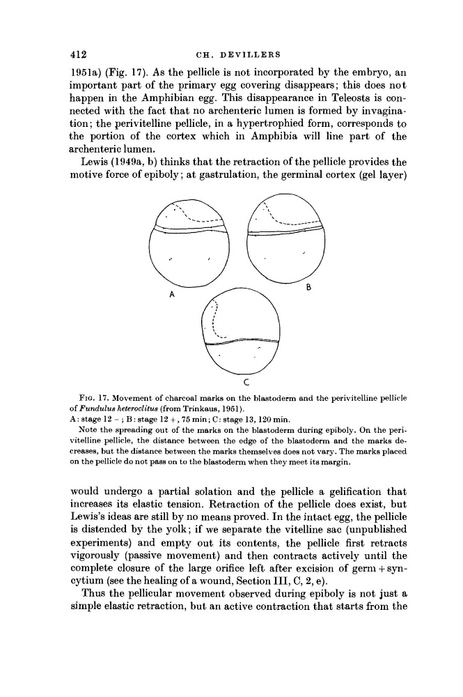
FIG. 17. Movement of charcoal marks on the blastoderm and the perivitelline pellicle
oí Fundulus heteroclitus (from Trinkaus, 1951).
A: stage 12 - ; Β: stage 12 +, 75min; C: stage 13, 120min.
Note the spreading out of the marks on the blastoderm during epiboly. On the perivitelline pellicle, the distance between the edge of the blastoderm and the marks decreases, but the distance between the marks themselves does not vary. The marks placed
on the pellicle do not pass on to the blastoderm when they meet its margin.
would undergo a partial solation and the pellicle a gelification that
increases its elastic tension. Retraction of the pellicle does exist, but
Lewis's ideas are still by no means proved. In the intact egg, the pellicle
is distended by the yolk; if we separate the vitelline sac (unpublished
experiments) and empty out its contents, the pellicle first retracts
vigorously (passive movement) and then contracts actively until the
complete closure of the large orifice left after excision of germ + syncytium (see the healing of a wound, Section III, C, 2, e).
Thus the pellicular movement observed during epiboly is not just a
simple elastic retraction, but an active contraction that starts from the
1951a) (Fig. 17). As the pellicle is not incorporated by the embryo, an
important part of the primary egg covering disappears; this does not
happen in the Amphibian egg. This disappearance in Teleosts is connected with the fact that no archenteric lumen is formed by invagination; the perivitelline pellicle, in a hypertrophied form, corresponds to
the portion of the cortex which in Amphibia will line part of the
archenteric lumen.
Lewis (1949a, b) thinks that the retraction of the pellicle provides the
motive force of epiboly; at gastrulation, the germinal cortex (gel layer)
CH. DEVILLERS
C
FIG. 17. Movement of charcoal marks on the blastoderm and the perivitelline pellicle
oí Fundulus heteroclitus (from Trinkaus, 1951).
A: stage 12 - ; Β: stage 12 +, 75min; C: stage 13, 120min.
Note the spreading out of the marks on the blastoderm during epiboly. On the perivitelline pellicle, the distance between the edge of the blastoderm and the marks decreases, but the distance between the marks themselves does not vary. The marks placed
on the pellicle do not pass on to the blastoderm when they meet its margin.
would undergo a partial solation and the pellicle a gelification that
increases its elastic tension. Retraction of the pellicle does exist, but
Lewis's ideas are still by no means proved. In the intact egg, the pellicle
is distended by the yolk; if we separate the vitelline sac (unpublished
experiments) and empty out its contents, the pellicle first retracts
vigorously (passive movement) and then contracts actively until the
complete closure of the large orifice left after excision of germ + syncytium (see the healing of a wound, Section III, C, 2, e).
Thus the pellicular movement observed during epiboly is not just a
simple elastic retraction, but an active contraction that starts from the
1951a) (Fig. 17). As the pellicle is not incorporated by the embryo, an
important part of the primary egg covering disappears; this does not
happen in the Amphibian egg. This disappearance in Teleosts is connected with the fact that no archenteric lumen is formed by invagination; the perivitelline pellicle, in a hypertrophied form, corresponds to
the portion of the cortex which in Amphibia will line part of the
archenteric lumen.
Lewis (1949a, b) thinks that the retraction of the pellicle provides the
motive force of epiboly; at gastrulation, the germinal cortex (gel layer)