

278
CHARLES
Ε.
WILDE
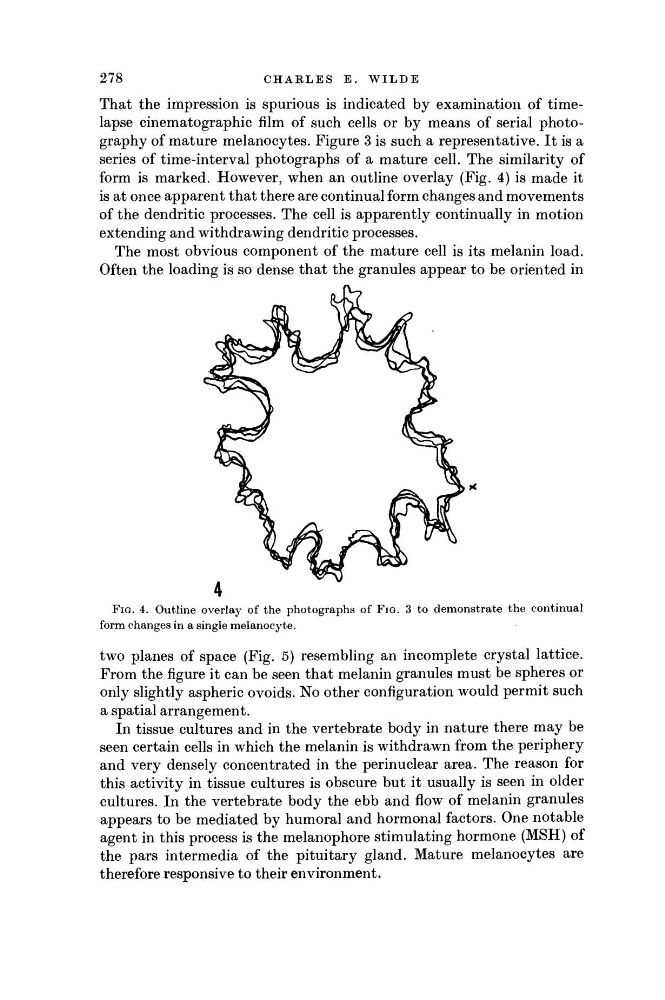
FIG. 4. Outline overlay of the photographs of FIG. 3 to demonstrate the continual
form changes in a single melanocyte.
two planes of space (Fig. 5) resembling an incomplete crystal lattice.
From the figure it can be seen that melanin granules must be spheres or
only slightly aspheric o voids. No other configuration would permit such
a spatial arrangement.
In tissue cultures and in the vertebrate body in nature there may be
seen certain cells in which the melanin is withdrawn from the periphery
and very densely concentrated in the perinuclear area. The reason for
this activity in tissue cultures is obscure but it usually is seen in older
cultures. In the vertebrate body the ebb and flow of melanin granules
appears to be mediated by humoral and hormonal factors. One notable
agent in this process is the melanophore stimulating hormone (MSH) of
the pars intermedia of the pituitary gland. Mature melanocytes are
therefore responsive to their environment.
That the impression is spurious is indicated by examination of timelapse cinematographic film of such cells or by means of serial photography of mature melanocytes. Figure 3 is such a representative. It is a
series of time-interval photographs of a mature cell. The similarity of
form is marked. However, when an outline overlay (Fig. 4) is made it
is at once apparent that there are continual form changes and movements
of the dendritic processes. The cell is apparently continually in motion
extending and withdrawing dendritic processes.
The most obvious component of the mature cell is its melanin load.
Often the loading is so dense that the granules appear to be oriented in
CHARLES
Ε.
WILDE
FIG. 4. Outline overlay of the photographs of FIG. 3 to demonstrate the continual
form changes in a single melanocyte.
two planes of space (Fig. 5) resembling an incomplete crystal lattice.
From the figure it can be seen that melanin granules must be spheres or
only slightly aspheric o voids. No other configuration would permit such
a spatial arrangement.
In tissue cultures and in the vertebrate body in nature there may be
seen certain cells in which the melanin is withdrawn from the periphery
and very densely concentrated in the perinuclear area. The reason for
this activity in tissue cultures is obscure but it usually is seen in older
cultures. In the vertebrate body the ebb and flow of melanin granules
appears to be mediated by humoral and hormonal factors. One notable
agent in this process is the melanophore stimulating hormone (MSH) of
the pars intermedia of the pituitary gland. Mature melanocytes are
therefore responsive to their environment.
That the impression is spurious is indicated by examination of timelapse cinematographic film of such cells or by means of serial photography of mature melanocytes. Figure 3 is such a representative. It is a
series of time-interval photographs of a mature cell. The similarity of
form is marked. However, when an outline overlay (Fig. 4) is made it
is at once apparent that there are continual form changes and movements
of the dendritic processes. The cell is apparently continually in motion
extending and withdrawing dendritic processes.
The most obvious component of the mature cell is its melanin load.
Often the loading is so dense that the granules appear to be oriented in