

168
F.
Ε.
LEHMANN
in E 9 , colchicine and water (test of regeneration under the action of the
combined solutions). The following tissue pieces were sampled on each
day of measurement: the regenerates, the adjoining 2 mm of stump
tissue and also the corresponding 2 mm slices from non-amputated
animals in test solution and in water (treated and untreated controls).
10 mm
07
1-0
I 0-5
O
U
Normal regeneration
5
10
15
Days after amputation
20
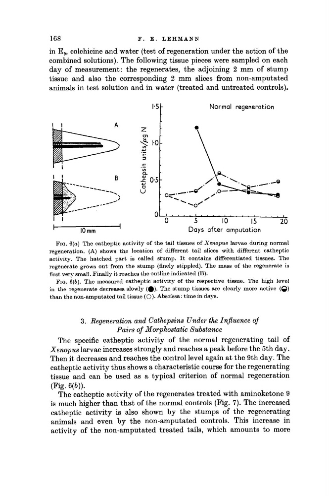
FIG. 6(a) The catheptic activity of the tail tissues of Xenopus larvae during normal
regeneration. (A) shows the location of different tail slices with different catheptic
activity. The hatched part is called stump. It contains differentiated tissues. The
regenerate grows out from the stump (finely stippled). The mass of the regenerate is
first very small. Finally it reaches the outline indicated (B).
FIG. 6(6). The measured catheptic activity of the respective tissue. The high level
in the regenerate decreases slowly (φ). The stump tissues are clearly more active (Q)
than the non-amputated tail tissue (O )· Abscissa: time in days.
3. Regeneration and Cathepsins Under the Influence of
Pairs of Morphostatic Substance
The specific catheptic activity of the normal regenerating tail of
Xenopus larvae increases strongly and reaches a peak before the 5th day.
Then it decreases and reaches the control level again at the 9th day. The
catheptic activity thus shows a characteristic course for the regenerating
tissue and can be used as a typical criterion of normal regeneration
(Fig. 6(6)).
The catheptic activity of the regenerates treated with aminoketone 9
is much higher than that of the normal controls (Fig. 7). The increased
catheptic activity is also shown by the stumps of the regenerating
animals and even by the non-amputated controls. This increase in
activity of the non-amputated treated tails, which amounts to more
F.
Ε.
LEHMANN
in E 9 , colchicine and water (test of regeneration under the action of the
combined solutions). The following tissue pieces were sampled on each
day of measurement: the regenerates, the adjoining 2 mm of stump
tissue and also the corresponding 2 mm slices from non-amputated
animals in test solution and in water (treated and untreated controls).
10 mm
07
1-0
I 0-5
O
U
Normal regeneration
5
10
15
Days after amputation
20
FIG. 6(a) The catheptic activity of the tail tissues of Xenopus larvae during normal
regeneration. (A) shows the location of different tail slices with different catheptic
activity. The hatched part is called stump. It contains differentiated tissues. The
regenerate grows out from the stump (finely stippled). The mass of the regenerate is
first very small. Finally it reaches the outline indicated (B).
FIG. 6(6). The measured catheptic activity of the respective tissue. The high level
in the regenerate decreases slowly (φ). The stump tissues are clearly more active (Q)
than the non-amputated tail tissue (O )· Abscissa: time in days.
3. Regeneration and Cathepsins Under the Influence of
Pairs of Morphostatic Substance
The specific catheptic activity of the normal regenerating tail of
Xenopus larvae increases strongly and reaches a peak before the 5th day.
Then it decreases and reaches the control level again at the 9th day. The
catheptic activity thus shows a characteristic course for the regenerating
tissue and can be used as a typical criterion of normal regeneration
(Fig. 6(6)).
The catheptic activity of the regenerates treated with aminoketone 9
is much higher than that of the normal controls (Fig. 7). The increased
catheptic activity is also shown by the stumps of the regenerating
animals and even by the non-amputated controls. This increase in
activity of the non-amputated treated tails, which amounts to more