

238
JOHN RUNNSTRÖM
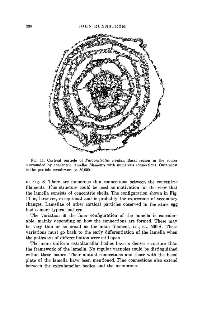
FIG. 11. Cortical particle of Paracentrotus lividus. Basal region in the center
surrounded by concentric lamellar filaments with numerous connections. Outermost
is the particle membrane, χ 80,000.
in Fig. 8. There are numerous thin connections between the concentric
filaments. This structure could be used as motivation for the view that
the lamella consists of concentric shells. The configuration shown in Fig.
11 is, however, exceptional and is probably the expression of secondary
changes. Lamellae of other cortical particles observed in the same egg
had a more typical pattern.
The variation in the finer configuration of the lamella is considerable, mainly depending on how the connections are formed. These may
be very thin or as broad as the main filament, i.e., ca. 500 A. These
variations must go back to the early differentiation of the lamella when
the pathways of differentiation were still open.
The more uniform extralamellar bodies have a denser structure than
the framework of the lamella. No regular vacuoles could be distinguished
within these bodies. Their mutual connections and those with the basal
plate of the lamella have been mentioned. Fine connections also extend
between the extralamellar bodies and the membrane.
JOHN RUNNSTRÖM
FIG. 11. Cortical particle of Paracentrotus lividus. Basal region in the center
surrounded by concentric lamellar filaments with numerous connections. Outermost
is the particle membrane, χ 80,000.
in Fig. 8. There are numerous thin connections between the concentric
filaments. This structure could be used as motivation for the view that
the lamella consists of concentric shells. The configuration shown in Fig.
11 is, however, exceptional and is probably the expression of secondary
changes. Lamellae of other cortical particles observed in the same egg
had a more typical pattern.
The variation in the finer configuration of the lamella is considerable, mainly depending on how the connections are formed. These may
be very thin or as broad as the main filament, i.e., ca. 500 A. These
variations must go back to the early differentiation of the lamella when
the pathways of differentiation were still open.
The more uniform extralamellar bodies have a denser structure than
the framework of the lamella. No regular vacuoles could be distinguished
within these bodies. Their mutual connections and those with the basal
plate of the lamella have been mentioned. Fine connections also extend
between the extralamellar bodies and the membrane.