

V I T E L L I N E M E M B R A N E A N D C O R T I C A L P A R T I C L E S 235
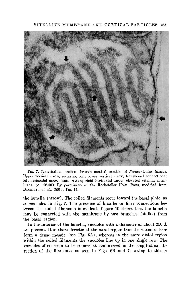
FIG. 7. Longitudinal section through cortical particle of Paracentrotus
lividus.
Upper vertical arrow, recurring coil; lower vertical arrow, transversal connections;
left horizontal arrow, basal region; right horizontal arrow, elevated vitelline membrane. X 105,000. By permission of the Rockefeller Univ. Press, modified from
Baxandall et al, 1964b, Fig. 14.)
the lamella (arrow). The coiled filaments recur toward the basal plate, as
is seen also in Fig. 7. The presence of broader or finer connections between the coiled filaments is evident. Figure 10 shows t h a t the lamella
may be connected with the membrane by two branches (stalks) from
the basal region.
In the interior of the lamella, vacuoles with a diameter of about 250 Â
are present. I t is characteristic of the basal region t h a t the vacuoles here
form a dense mosaic (see Fig. 6A), whereas in the more distal region
within the coiled filaments the vacuoles line up in one single row. The
vacuoles often seem to be somewhat compressed in the longitudinal direction of the filaments, as seen in Figs. 6B and 7; owing to this, a
FIG. 7. Longitudinal section through cortical particle of Paracentrotus
lividus.
Upper vertical arrow, recurring coil; lower vertical arrow, transversal connections;
left horizontal arrow, basal region; right horizontal arrow, elevated vitelline membrane. X 105,000. By permission of the Rockefeller Univ. Press, modified from
Baxandall et al, 1964b, Fig. 14.)
the lamella (arrow). The coiled filaments recur toward the basal plate, as
is seen also in Fig. 7. The presence of broader or finer connections between the coiled filaments is evident. Figure 10 shows t h a t the lamella
may be connected with the membrane by two branches (stalks) from
the basal region.
In the interior of the lamella, vacuoles with a diameter of about 250 Â
are present. I t is characteristic of the basal region t h a t the vacuoles here
form a dense mosaic (see Fig. 6A), whereas in the more distal region
within the coiled filaments the vacuoles line up in one single row. The
vacuoles often seem to be somewhat compressed in the longitudinal direction of the filaments, as seen in Figs. 6B and 7; owing to this, a