

234
J O H N R U N N S T R Ö M
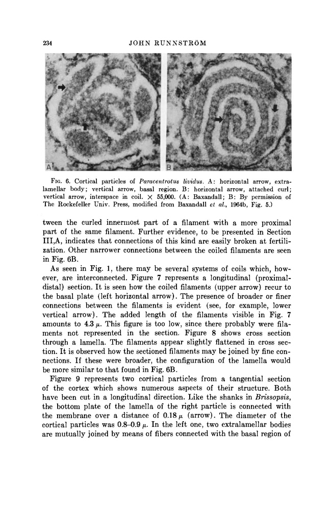
FIG. 6. Cortical particles of Paracentrotus lividus. A: horizontal arrow, extralamellar body; vertical arrow, basal region. B: horizontal arrow, attached curl;
vertical arrow, interspace in coil. X 55,000. (A: Baxandall; B: By permission of
The Rockefeller Univ. Press, modified from Baxandall et al., 1964b, Fig. 5.)
tween the curled innermost p a r t of a filament with a more proximal
p a r t of the same filament. Further evidence, to be presented in Section
ΙΙΙ,Α, indicates t h a t connections of this kind are easily broken a t fertilization. Other narrower connections between the coiled filaments are seen
in Fig. 6B.
As seen in Fig. 1, there may be several systems of coils which, however, are interconnected. Figure 7 represents a longitudinal (proximaldistal) section. I t is seen how the coiled filaments (upper arrow) recur to
the basal plate (left horizontal arrow). The presence of broader or finer
connections between the filaments is evident (see, for example, lower
vertical arrow). The added length of the filaments visible in Fig. 7
amounts to 4.3 μ. This figure is too low, since there probably were filaments not represented in the section. Figure 8 shows cross section
through a lamella. The filaments appear slightly flattened in cross section. I t is observed how the sectioned filaments m a y be joined by fine connections. If these were broader, the configuration of the lamella would
be more similar to t h a t found in Fig. 6B.
Figure 9 represents two cortical particles from a tangential section
of the cortex which shows numerous aspects of their structure. Both
have been cut in a longitudinal direction. Like the shanks in Brissopsis,
the bottom plate of the lamella of the right particle is connected with
the membrane over a distance of 0.18 μ (arrow). The diameter of the
cortical particles was 0.8-0.9 μ. In the left one, two extralamellar bodies
are mutually joined by means of fibers connected with the basal region of
J O H N R U N N S T R Ö M
FIG. 6. Cortical particles of Paracentrotus lividus. A: horizontal arrow, extralamellar body; vertical arrow, basal region. B: horizontal arrow, attached curl;
vertical arrow, interspace in coil. X 55,000. (A: Baxandall; B: By permission of
The Rockefeller Univ. Press, modified from Baxandall et al., 1964b, Fig. 5.)
tween the curled innermost p a r t of a filament with a more proximal
p a r t of the same filament. Further evidence, to be presented in Section
ΙΙΙ,Α, indicates t h a t connections of this kind are easily broken a t fertilization. Other narrower connections between the coiled filaments are seen
in Fig. 6B.
As seen in Fig. 1, there may be several systems of coils which, however, are interconnected. Figure 7 represents a longitudinal (proximaldistal) section. I t is seen how the coiled filaments (upper arrow) recur to
the basal plate (left horizontal arrow). The presence of broader or finer
connections between the filaments is evident (see, for example, lower
vertical arrow). The added length of the filaments visible in Fig. 7
amounts to 4.3 μ. This figure is too low, since there probably were filaments not represented in the section. Figure 8 shows cross section
through a lamella. The filaments appear slightly flattened in cross section. I t is observed how the sectioned filaments m a y be joined by fine connections. If these were broader, the configuration of the lamella would
be more similar to t h a t found in Fig. 6B.
Figure 9 represents two cortical particles from a tangential section
of the cortex which shows numerous aspects of their structure. Both
have been cut in a longitudinal direction. Like the shanks in Brissopsis,
the bottom plate of the lamella of the right particle is connected with
the membrane over a distance of 0.18 μ (arrow). The diameter of the
cortical particles was 0.8-0.9 μ. In the left one, two extralamellar bodies
are mutually joined by means of fibers connected with the basal region of