

ULTRASTRUCTURE OF DEVELOPING AMPHIBIAN EGG
161
3. Cortical Granules
The presence in anuran oocytes of a layer of cortical granules close to
the plasmalemma was first seen with the electron microscope by Kemp
(1953). Later, Wartenberg and Gusek (1960) confirmed their presence in
the subcortical region and also found them distributed in deeper parts of the
cytoplasm.
The cortical granules, as seen with the electron microscope, are spherical
masses composed of a finely granular homogeneous material having a dense
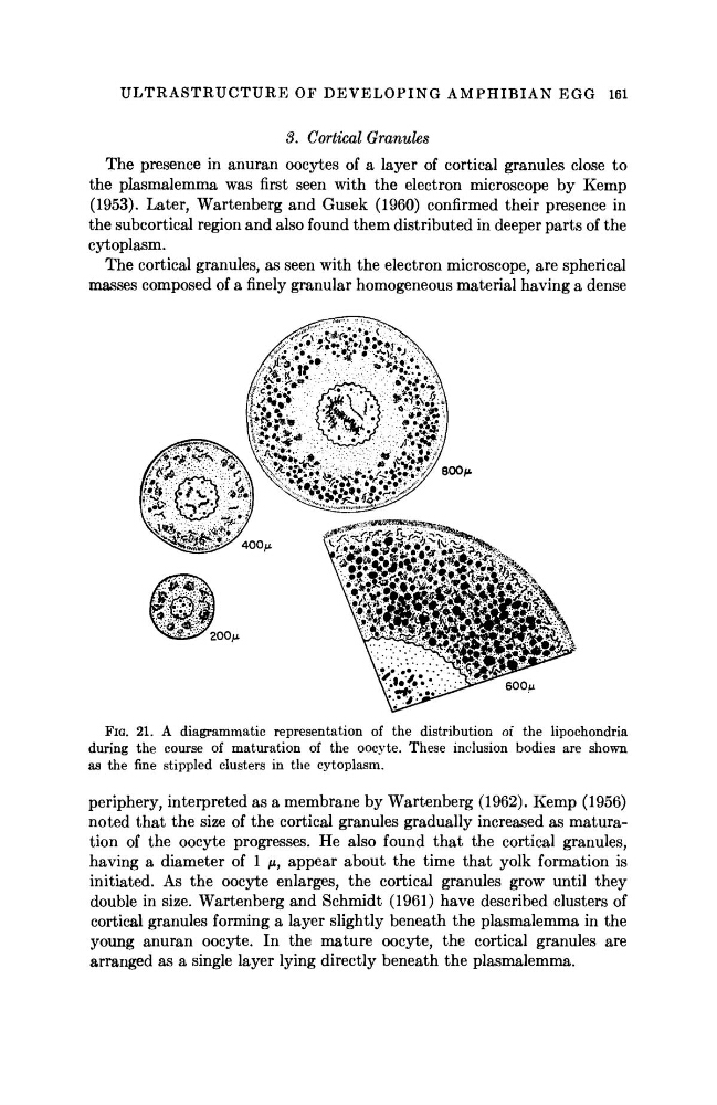
FIG. 21. A diagrammatic representation of the distribution of the lipochondria
during the course of maturation of the oocyte. These inclusion bodies are shown
as the fine stippled clusters in the cytoplasm.
periphery, interpreted as a membrane by Wartenberg (1962). Kemp (1956)
noted that the size of the cortical granules gradually increased as maturation of the oocyte progresses. He also found that the cortical granules,
having a diameter of 1 μ, appear about the time that yolk formation is
initiated. As the oocyte enlarges, the cortical granules grow until they
double in size. Wartenberg and Schmidt (1961) have described clusters of
cortical granules forming a layer slightly beneath the plasmalemma in the
young anuran oocyte. In the mature oocyte, the cortical granules are
arranged as a single layer lying directly beneath the plasmalemma.
161
3. Cortical Granules
The presence in anuran oocytes of a layer of cortical granules close to
the plasmalemma was first seen with the electron microscope by Kemp
(1953). Later, Wartenberg and Gusek (1960) confirmed their presence in
the subcortical region and also found them distributed in deeper parts of the
cytoplasm.
The cortical granules, as seen with the electron microscope, are spherical
masses composed of a finely granular homogeneous material having a dense
FIG. 21. A diagrammatic representation of the distribution of the lipochondria
during the course of maturation of the oocyte. These inclusion bodies are shown
as the fine stippled clusters in the cytoplasm.
periphery, interpreted as a membrane by Wartenberg (1962). Kemp (1956)
noted that the size of the cortical granules gradually increased as maturation of the oocyte progresses. He also found that the cortical granules,
having a diameter of 1 μ, appear about the time that yolk formation is
initiated. As the oocyte enlarges, the cortical granules grow until they
double in size. Wartenberg and Schmidt (1961) have described clusters of
cortical granules forming a layer slightly beneath the plasmalemma in the
young anuran oocyte. In the mature oocyte, the cortical granules are
arranged as a single layer lying directly beneath the plasmalemma.