

160
S A U L W I S C H N I T Z E R
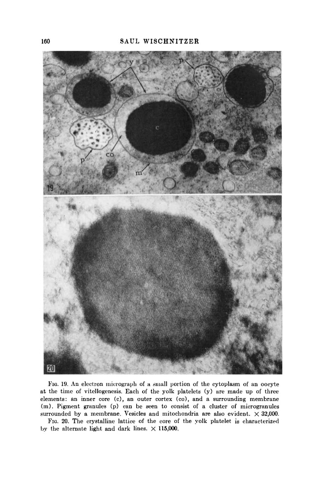
FIG. 19. An electron micrograph of a small portion of the cytoplasm of an oocyte
at the time of vitellogenesis. Each of the yolk platelets (y) are made up of three
elements: an inner core (c), an outer cortex (co), and a surrounding membrane
(m). Pigment granules (p) can be seen to consist of a cluster of microgranules
surrounded by a membrane. Vesicles and mitochondria are also evident. X 32,000.
FIG. 20. The crystalline lattice of the core of the yolk platelet is characterized
by the alternate light and dark lines. X 115,000.
S A U L W I S C H N I T Z E R
FIG. 19. An electron micrograph of a small portion of the cytoplasm of an oocyte
at the time of vitellogenesis. Each of the yolk platelets (y) are made up of three
elements: an inner core (c), an outer cortex (co), and a surrounding membrane
(m). Pigment granules (p) can be seen to consist of a cluster of microgranules
surrounded by a membrane. Vesicles and mitochondria are also evident. X 32,000.
FIG. 20. The crystalline lattice of the core of the yolk platelet is characterized
by the alternate light and dark lines. X 115,000.